Pinna, or Auricle
The pinna, or auricle, is a highly visible structure. Carriage of the pinna is breedspecific in the dog but mostly upright in the cat. It is designed to localize and collect sound waves and transmit them to the tympanic membrane (eardrum).
The ear is moved by three sets of muscles (rostral, ventral, and caudal) that are innervated by branches of the facial nerve (cranial nerve VII).The leaf-shaped pinna of the external ear is broad with medial (rostral) and lateral (caudal) margins. The caudal margin of the pinna exhibits a cutaneous pouch called the marginal pouch (Figure 1-3). This pouch has no obvious function. The skin on the concave surface of the pinna is very tightly connected to the underlying auricular cartilage, accentuating all the auricular prominences (see Figure 1-3). The skin covering the auricular cartilage may show breed-specific pigmentation. The shape and size of the external ear vary greatly among different breeds of dogs, mainly owing to the auricular cartilage that forms the skeleton of the pinna. It is the largest cartilage of the external ear. The broad auricular cartilage has numerous holes (see Figure 1-1), which are traversed by branches arising from the caudal auricular artery.
The auricular cartilage is broad dorsally and funnels to a narrow tubelike structure, the tubus auris, which fits around the annular cartilage ring. The parotid salivary gland occupies the base of the external ear, partially surrounding the tubus auris (Figures 1-4 and 1-5). The tubus auris encloses the vertical part of the external canal and, together
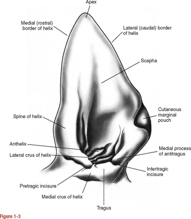
Anatomical features of the left external ear of the dog.
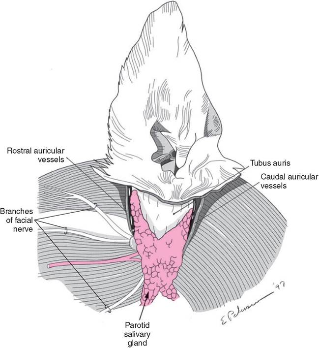
Figure 1-4
Relationships of the tubus auris to the parotid salivary gland and auricular vessels.
The facial nerve runs deep to the parotid salivary gland, immediately ventral to the annular cartilage, and gives off motor branches to the facial muscles.with the tragal, antitragal, and antihelicene borders, forms the external acoustic meatus (see Figure 1-3).
Usually, the entrance to the external ear canal is guarded by a few fine hairs. Certain breeds, such as Airedales and Old English sheepdogs, exhibit hairy external ear canals. The external ear canal of the cat is devoid of hairs, and the ear canal is well ventilated. This may be a significant factor contributing to the lower incidence of external ear canal infections in cats. A hairy ear canal interferes with proper drainage and aeration of the canal in chronic otitis externa complicated by granulomatous lesions, leading to exacerbation of the condition.
Rights were not granted to include this figure in electronic media. Please refer to the printed publication.
Figure 1-5
Structure of the ear canal. (Redrawn from Bojrab MJ: Current techniques in small animal surgery I, Philadelphia, 1975, Lea & Febiger.)
Annular Cartilage
The annular cartilage is part of the external ear canal. The pinna, formed by the coneshaped auricular cartilage, articulates with the annular cartilage. The annular cartilage is a ring-shaped structure attached to the bony orbit of the external acoustic meatus of the temporal bone. A tubular cartilage piece, the annular cartilage surrounds the osseous external acoustic meatus (see Figure 1-7). It is attached to the bony rim of the external acoustic meatus by fibrous tissue that permits some degree of movement of the external ear. The annular cartilage encloses the horizontal part of the external ear canal.
Scutiform Cartilage
The scutiform cartilage is an L-shaped structure located over the temporalis muscle. It does not contribute to the formation of the external ear or its canal. The scutiform cartilage is attached to the midline raphe of the head and neck by numerous muscles (see Figure 1-2). Muscles also extend from the scutiform cartilage to the auricular cartilage. The scutiform cartilage functions like a fulcrum, providing for efficient movement of the auricle. It can be considered to function like a sesamoid cartilage. It lies over a fat cushion (corpus adiposum auriculae) on the dorsal surface of the temporalis muscle.
More on the topic Pinna, or Auricle:
- Pinna Anatomy
- Dermatoses of the Concave Pinna
- Neoplasia of the Pinna
- 12 Diseases that Affect th e Pinna
- Most skin diseases may affect the pinna in dogs and cats, but other parts of the body can also be involved.
- Dermatoses of the Ear Margin
- Aural Hematoma
- Atopic Dermatitis
- Insect Bite Dermatitis
- Sarcoptic Mange
- Cosmetic and Therapeutic Otoplasty
- Atrial Thrombosis
- Pruritic Dermatoses
- Contact Dermatitis