Atopic Dermatitis
Erythema of the concave pinna is seen in more than 50% of atopic dermatitis cases in dogs and is the only symptom for a year or more before the development of other symptoms in at least 3% of canine cases.10-12 Associated pruritus is intense (Figure 12-4).
Lichenification and thickening of the concave pinna usually occur. Seborrhea and Malassezia dermatitis are common complications. This frequent feature might be explained in part by the observation made on 10 normal dogs, where mast cell counts were found to be highest in the medial and lateral pinna.7 Otitis externa, cheilitis, and conjunctivitis are also frequently encountered in canine atopic dermatitis and also cause pinnal pruritus and trauma.
The clinical presentation of atopic dermatitis is very different in cats. Otitis externa and the related pinnal trauma caused by pruritus are uncommon, even rare. Atopic dermatitis is a common underlying cause of the initially non-lesional pruritus of the face, neck and pinna, even though Otodectes cynotis and food allergy are the main differential diagnosis for this condition in cats.
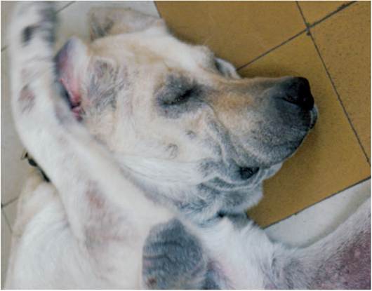
Figure 12-4
Intense pruritus of the inner pinnae in a 5-year-old female atopic Labrador with secondary Malassezia dermatitis and otitis externa.
More on the topic Atopic Dermatitis:
- Atopic Dermatitis
- Allergic Dermatitis
- Contact Dermatitis
- Staphylococcus aureus Infection: Ulcerative Dermatitis
- Insect Bite Dermatitis
- Bedding-Associated Dermatitis
- Pruritic Dermatoses
- ECZEMATOUS SKIN DISORDERS
- Most skin diseases may affect the pinna in dogs and cats, but other parts of the body can also be involved.
- Dermatoses of the Concave Pinna
- ORAL CAVITY INFLAMMATIONS
- Signalment and History
- Leporacarus gibbus Infestation
- Fusobacterium necrophorum Infection: Schmorl’s Disease, Necrobacillosis
- Corynebacterium bovis Infection: Coryneform Hyperkeratosis; Scaly Skin Disease
- OTHER CAUSES OF FOOT LAMENESS