Staphylococcus aureus Infection: Ulcerative Dermatitis
Staphylococcus aureus is a ubiquitous commensal bacterium that inhabits the skin and mucous membranes of laboratory rodents. Infection is usually subclinical, but S. aureus may be associated with ulcerative skin lesions among adult rats and occasionally vesicular dermatitis in infant rats.
The incidence of ulcerative dermatitis may vary from 1-2% to over 20% in certain populations of rats. The syndrome is particularly common in NK-defi- cient beige (Chediak-Higashi syndrome) rats. Lesions are most common in males. Trauma with persistent irritation appears to be an important contributing factor. Toenail clipping or amputation of the toes of the hind feet has resulted in remission of skin lesions, emphasizing the role of self-trauma in the disease. Changes ranging from alopecia to superficial ulceration have been associated with linoleic acid deficiency in Dahl rats. Linoleic acid plays a role in the cornification process and the maintenance of healthy skin. A marked increase in the numbers of S. aureus has been observed on the skin of mice deficient in linoleic acid. The significance, if any, of these findings in naturally occurring cases of ulcerative dermatitis in rats is unknown.Pathology
Irregular, circumscribed red ulcerative skin lesions occur over the shoulder and rib cage, submandibular regions,
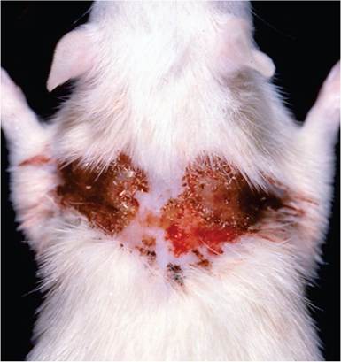
FIG. 2.41. Ulcerative dermatitis of the dorsal neck and interscapular regions in a Sprague-Dawley rat. This syndrome is associated with Staphylococcus aureus infection.
neck, ears, and head (Fig. 2.41). In acute cases, microscopic examination of lesions reveals an ulcerative dermatitis, with underlying coagulation necrosis of the underlying dermis. In adjacent areas, there is hyperplasia of the epidermis, with leukocytic infiltration into the underlying dermis (Fig.
2.42). Colonies of Gram-positive coccoid bacteria may be present within the proteinaceous material on the surface of lesions. There may be variable degeneration and leukocytic infiltration of the adnexae in affected areas. In lesions of some duration, dermal sclerosis and mononuclear cell infiltration are prominent features. Healed lesions frequently have dense collagenous tissue in the dermis, with loss of hair follicles and other adnexae. Histopathology suggests a role of epidermolytic toxin, as early stages are reminiscent of burn lesions. Lesions in deeper tissues may manifest botryomycotic features of the Splendore- Hoeppli response, with colonies of Gram-positive cocci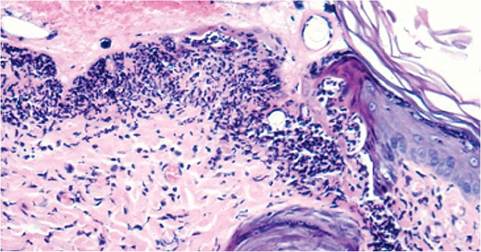
FIG. 2.42. Skin of a rat with ulcerative dermatitis. There is a sharply demarcated area of epidermal coagulation necrosis with underlying leukocytic infiltration. Discrete colonies of darkly staining coccoid bacteria are present within the surface exudate. (Source: T.W. Forest.)
surrounded by radiate eosinophilic material. Mastitis, subcutaneous abscesses, and infection of prepucial glands may also be associated with S. aureus infection.
Diagnosis
The presence of the typical ulcerative lesion with colonization by Gram-positive cocci is diagnostic. Bacterial cultures yield coagulase-positive S. aureus. Differential diagnoses include mycotic infections, fighting injuries, and, rarely, skin lesions associated with epitheliotropic lymphoid tumors (mycosis fungoides).
More on the topic Staphylococcus aureus Infection: Ulcerative Dermatitis:
- Staphylococcus spp. Infections
- STAPHYLOCOCCUS INFECTIONS
- Sexually Transmitted Infections: Ulcerative Diseases
- Allergic Dermatitis
- Atopic Dermatitis
- Contact Dermatitis
- Insect Bite Dermatitis
- Bedding-Associated Dermatitis
- Atopic Dermatitis
- Picornavirus Infection: Mouse Encephalomyelitis Virus Infection
- Arenavirus Infection: Lymphocytic Choriomeningitis Virus Infection
- Arterivirus Infection: Lactate Dehydrogenase-Elevating Virus Infection
- Coronavirus Infection: Mouse Hepatitis Virus Infection