Staphylococcus spp. Infections
Coagulase-negative staphylococci are commensal and opportunistic pathogenic bacteria that commonly inhabit the skin, nasopharynx, and intestine. In the mouse, disease has been associated with Staphylococcus aureus and S.
xylosus. Staphylococcus epidermidis and other species may be isolated from skin and mucous membranes, but have yet to be incriminated as pathogens in mice. Staphylococcus spp. are associated with several relatively distinct but overlapping common syndromes in mice:Chronic suppurative inflammation. Staphylococcus aureus is often associated with chronic suppurative inflammation of skin adnexae, conjunctiva, periorbital tissue, preputial glands, and regional lymph nodes. Euthymic mice, as well as urokinase plasminogen activator-deficient mice, may develop suppurative conjunctivitis, ophthalmitis, and periocular abscessation, with involvement of regional lymph nodes. Ocular lesions tend to occur in strains of mice that are prone to development of conjunctivitis or with ocular defects, such as BALB and B6 mice. Preputial gland inflammation and abscesses are sporadically common in male mice. Occasionally, deep visceral abscesses may also be found. Lymphadenitis and abscesses have botryomy- cotic-like features, characterized by necrotizing suppurative inflammation with a central core of necrotic neutrophils and colonies of Gram-positive bacteria surrounded by bright eosinophilic amorphous to fibrillar Splendore-Hoeppli material (Fig. 1.76). The authors have also observed botryomycotic abscesses involving bone and extending into the nasal cavity associated with foreign body periodontitis.
Furunculosis. Distorted vibrissal hair shaft growth and impaired T-cell function predispose athymic nude mice to staphylococcal furunculosis of the muzzle (Fig. 1.77). These mice frequently have suppurative lymphadenitis of the regional lymph nodes.
Microscopic findings are similar to those described above with botryomycotic features.Ulcerative dermatitis. An important form of staphylococcal disease is ulcerative dermatitis in association with S. aureus or S. xylosus. Staphylococci produce an array of biologically active proteins, including
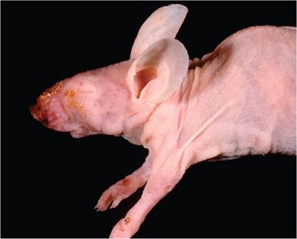
FIG. 1.77. Muzzle furunculosis in a nude mouse associated with Staphylococcus aureus infection.
hemolysins, nucleases, proteases, lipases, hyaluronidase, and collagenase, and many produce exotoxins, including exfoliative toxins, leukocidin, and several superantigens, particularly enterotoxin A, enterotoxin B, enterotoxin C, and toxic shock syndrome toxin-1. This armamentarium is important in understanding the pathogenesis of staphylococcal ulcerative dermatitis in mice (and rats), which is characterized by superficial colonization of bacteria on the surface of the skin, with underlying burn-like features.
Ulcerative dermatitis is quite common in young and aged B6, BALB/c, DBA/2, and C3H/He mice and their hybrids, as well as other strains and stocks of mice. Disease prevalence is linked to other predisposing factors, including behavioral idiosyncrasies and ectoparasite hypersensitivity. For example, B6 mice are prone to trichotillomania and frequently suffer from ulcerative dermatitis (Fig. 1.78). Superficial excoriation of the skin is followed by colonization with Staphylococcus, pruritus, and then development of progressive necrotizing dermatitis. The syndrome has been well studied in hairless DS-Nh mice infected with S. aureus. DS-Nh mice normally manifest excessive scratching behavior. When they are housed in a conventional environment, they develop erythema, excoriation, and erosion around their head and neck. When first introduced to the environment, the mice become colonized with several Staphylococcus species, but S. aureus gradually becomes the dominant skin commensal over time, with selection for S. aureus types that produce enterotoxin C.
Lesions do not develop in mice maintained under Staphylococcus-free barrier conditions. A similar syndrome is associated with S. xylosus in athymic nude mice and B6-Nos2tm1Lau (NOS2) mice, which have defective inducible NO synthase. NOS2 mice have less severe disease compared to nude mice, which develop more severe disease with mortality. Notably, skin lesions do not appear to be extensive enough in nude mice to explain the high mortality, suggesting that the mice are succumbing to systemic toxic events. Immunocompetent SJL mice (which are NK cell-deficient) have also been reported to develop necrotic dermatitis on their tails in association with S. xylosus. Severe ulcerative conjunctivitis and dermatitis, with extensive facial and neck involvement, occur in CD18 (leukocyte integrin) null mice on B6 and 129 mixed background. The Staphylococcus isolate was not speciated, but lesions contained prominent colonies of staphylococcal-type bacteria in the superficial exudate, consistent with better characterized forms of this disease.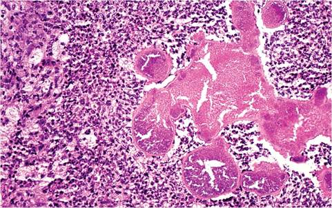
FIG. 1.76. Cervical lymph node, illustrating the botryomycotic inflammatory response due to Staphylococcus aureus infection. There are bacterial colonies and central aggregates of eosinophilic material consistent with a Splendore-Hoeppli reaction.
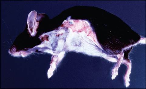
FIG. 1.78. Ulcerative dermatitis in a B6 mouse associated with Staphylococcus aureus infection. Note that the ventral abdomen is bald due to trichotillomania, with ulcerative dermatitis on the side.
Staphylococcal ulcerative dermatitis is characterized by focal small to large chronic ulcerative lesions, often around the head and neck, but also on the trunk or base of the tail. In the tail form of disease, gangrene and sloughing of the tail may occur. Apparently, the location of the skin lesions is associated with the initiating cause of excoriation (ectoparasites, grooming, fighting, etc.).
Regardless of location, the microscopic appearance of these lesions is similar (Fig. 1.79), with prominent colonies of Gram-positive organisms in the superficial exudate, and acute coagulative necrosis of the underlying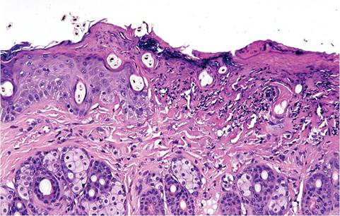
FIG. 1.79. Skin from a mouse with ulcerative dermatitis. Note the coagulation necrosis of the epidermis and dermis with the presence of bacterial colonies on the surface.
epidermis and dermis, which can extend to the subcutis and panniculus carnosis. The lesions are reminiscent of 1st, 2nd, or 3rd degree burns, which implicate the toxins elaborated by the Staphylococcus colonizing the surface of the lesions. Varying degrees of leukocytic infiltration and granulation are also present. The chronic lesions of Staphyloccal disease result in acceleration of multi- systemic amyloidosis and splenomegaly, with marked extramedullary myelopoiesis in liver and spleen. Plas- macytosis is present in regional lymph nodes. Scaly skin disease in nude mice, similar to the disease caused by C. bovis, has been associated with S. xylosus. However, lesions were more inflammatory and included foci of epidermal ulceration and pustule formation.
Diagnosis
An etiologic diagnosis requires isolation and speciation, but factors that predispose to susceptibility should be investigated, including immune status, strain-related behavior patterns, ectoparasitism, and ectoparasite- related hypersensitivity. Differential diagnoses include abscessation due to other bacterial infections and streptococcal necrotizing dermatitis. Amputation of the tail secondary to Staphylococcus-related dry gangrene must be differentiated from mousepox and tail lesions in mice infected with Mycobacterium chelonae.
More on the topic Staphylococcus spp. Infections:
- STAPHYLOCOCCUS INFECTIONS
- Other Bacterial Infections Corynebacterium spp. Infections
- Helicobacter spp. Infections
- Helicobacter spp. Infections
- Mycobacterium spp. Infections
- Bacterial Enteric Infections Brachyspira spp. Infection
- Cryptosporidium spp. Infections
- Mycoplasma spp. Infections
- Staphylococcus aureus Infection: Ulcerative Dermatitis
- Using PCR and 16S rRNA sequence analysis, gastric biopsy specimens were evaluated for the presence of Helicobacter spp. among pet, laboratory, and commercial rabbits. Rabbits from all sources tested positive for Helicobacter spp. Most of th
- Malassezia spp. Infection: Malasseziasis
- Helicobacter spp. Infection
- Aspergillus spp. Infection