Mycoplasma spp. Infections
Mycoplasmae are pleomorphic organisms that lack a cell wall and are enclosed by a single limiting membrane. Despite their lack of a cell wall, they are genetically related to several Gram-positive bacteria (Streptococcus, Lactobacillus, Clostridium, etc.).
Their lack of a cell wall places them within the class Mollicutes. Mice are host to several Mycoplasma species, which cluster into two groups, the “pneumoniae” and “hemotropic” groups. With the exception of Mycoplasma pulmonis, members of the pneumoniae group are marginally pathogenic or nonpathogenic and inhabit not only the respiratory tract but also the genital tract. Sequence analysis of the 16S rRNA genes has revealed that the genera Epery- throzoon and Hemobartonella, previously considered Rickettsiae, are hemotropic mycoplasmas (often referred to as hemoplasmas) that infect erythrocytes. These include Mycoplasma coccoides (formerly Eperythrozoon coccoides) and Mycoplasma haemomuris (formerly Hemobartonella muris).Respiratory and Genital Mycoplasmosis
Laboratory mice are host to several Mycoplasma species within the pneumoniae group, including M. pulmonis, M. arthritidis, M. neurolyticum, M. collis, and M. muris. An unclassified Mycoplasma, the “gray lung” agent, appears to be distantly related to M. pulmonis (84% homology) and more closely related to M. hominis (94% homology). The name M. ravipulmonis (ravi, gray; pulmonis, lung) has been proposed. Mycoplasma pulmonis, M. arthritidis, and M. neurolyticum inhabit the upper respiratory tract, and M. collis and M. muris inhabit the genital tract. Only M. pulmonis is a significant natural pathogen that produces respiratory and genital tract disease in mice and rats. Mycoplasma arthritidis may cause respiratory disease following intranasal inoculation, but under natural conditions, it is generally nonpathogenic. It is problematic primarily because it can cause seroconversion to M.
pulmonis. Mycoplasma arthritidis may induce arthritis when inoculated intravenously, thereby earning its name, but M. pulmonis can induce arthritis naturally or following either intravenous or intranasal inoculation. Mycoplasma neurolyticum is the causative agent of “rolling disease,” a term used to denote the neurologic signs associated with the exotoxin that follows experimental intracerebral inoculation of the organism in mice. Spontaneous outbreaks of conjunctivitis have been associated with M. neurolyticum infection in young mice, but the organism is nonpathogenic under most conditions and is exceedingly rare or nonexistent among laboratory mice.Epizootiology and Pathogenesis
Prior to and during the 1960s, infections with M. pulmonis were widespread among colonies of laboratory mice. With improvements in management practices, there has been a marked reduction in the prevalence of infected colonies of mice. Exposure occurs by aerosol transmission, but infection may also be venereally transmitted. Newborn animals become infected during the first few weeks of life from contact with infected mothers. Transplacental transmission can occur in rats and is likely, but not documented, in immunodeficient mice with disseminated infections. Mycoplasma has the potential of being transmitted through cesarean transfer, embryo transfer, or in vitro fertilization.
Compared with the laboratory rat, mice are relatively resistant to disease, and experimental disease severity is closely linked to inoculum dose. Susceptibility to disease depends upon the strain or isolate of M. pulmonis, but also on the genetic strain of mouse. Genetic resistance is complex and does not appear to be H-2 linked. C57BR, B6, and B10 mice are resistant, whereas C57L, SJL, BALB, A/J, C3H/HeJ, C3H/HeN, C3HeB, SWR, AKR, CBA/N, C58, and DBA/2 mice have been shown to be variably susceptible. Most experimental studies have compared disease between susceptible C3H and resistant B6 mice, and female mice have been found to develop more severe disease.
Chronic suppurative arthritis has been produced in immunocompetent mice inoculated intravenously with M. pulmonis. Athymic nude, thymecto- mized, CBA/N (X-linked immunodeficient), and SCID mice inoculated intranasally with M. pulmonis develop significantly less severe respiratory disease compared to immunocompetent mice, but have disseminated infection with severe polyarthritis. Spontaneous cases of M. pulmonis associated arthritis have not been reported in mice, but the potential for this manifestation is real with the increased use of immunodeficient mice.Mycoplasma pulmonis colonizes the apical cell membranes of respiratory epithelium and interferes with mucociliary clearance. Mycoplasmosis is exacerbated by viral infections, particularly Sendai virus, by other bacteria, including Pasteurella pneumotropica, and by environmental ammonia levels. These cofactors probably play a significant role in driving subclinical mycoplasmal infections into overt disease. Mycoplasma pulmonis is mitogenic for B cells, which contributes to the pathology (peribronchiolar lymphoplasmacytic infiltrates) observed in the lungs. CAR bacillus has similar mechanisms of altered mucociliary clearance and B-cell mitogenesis, thereby causing similar disease. Coinfection with CAR bacillus in cases of mycoplasmosis is common. The acquired immune response is important in limiting hematogenous dissemination of the infection, but contributes little to elimination of infection or resolution of disease (for additional information, see Rat Chapter 2, “M. pulmonis infection”).
Pathology
Infection is often subclinical or mild in mice. In natural outbreaks of disease, affected mice may exhibit weight loss, dyspnea, and a characteristic “chattering” sound. Mice with otitis may display head tilt, circling, or vestibular signs. Mucopurulent exudate may be present in the nasal passages, tympanic bullae, trachea, and major airways. In advanced cases, anteroventral gray-purple areas of atelectasis, bronchiolectasis, bronchopneumonia, and raised yellow-tan nodules with abscessation may be evident on gross examination (Fig.
1.68). On microscopic examination, suppurative rhinitis, with neutrophil and lymphocyte infiltration and hyperplasia of submucosal glands, are characteristic findings. In the respiratory epithelium of the nasal passages and major airways, there may be loss of cilia and flattening of epithelial lining cells. In association with the chronic suppurative process, syncytia may be present in affected nasal mucosa and larynx (Fig. 1.69). Suppurative otitis media (Fig. 1.70) is frequently found, with extension to otitis interna and meningitis in some cases. In the lower respiratory tract, lesions vary from discrete peribronchiolar and perivascular lymphocytic and plasma cell infiltration to chronic suppurative bronchiolitis and alveolitis, with mobilization of alveolar macrophages. In advanced cases, there may be squamous metaplasia of respiratory epithelium, bronchiolectasis, and abscessation, with obliteration of the normal architecture. Mice do not develop the intense peribronchiolar lymphocytic infiltrates and severe bronchiolectasis that are common features of respiratory mycoplasmosis in the rat.Although more common in the rat, chronic suppurative oophoritis, perioophoritis, salpingitis, and
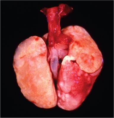
FIG. 1.68. Lungs from a mouse with respiratory mycoplasmosis. Note the multiple pale raised bronchiolectatic lesions.
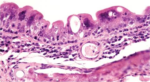
FIG. 1.69. Larynx from a mouse with chronic Mycoplasma pulmonis infection. Multinucleated giant cells are present in the respiratory epithelium. This is a common feature of the disease in this species that is not seen in rats.
endometritis are readily induced experimentally in mice and have been reported to occur in naturally infected mice. Isolation from the vagina and uterus of naturally infected mice and mice in contact with experimentally infected mice has also been documented.
Disseminated infection in SCID mice has been shown to result in suppurative splenitis, pericarditis, myocarditis, atrioventricular valvulitis, and polyarthritis. Although arthritis has not been described naturally, it is readily induced experimentally in B-cell-deficient, SCID, and C3H/HeN mice.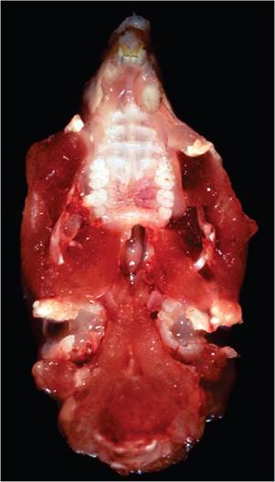
FIG. 1.70. Bilateral chronic suppurative otitis media due to Mycoplasma pulmonis infection. Note the presence of exudate in the opened tympanic bullae.
Diagnosis
Gross and microscopic lesions are characteristic. Histological assessment should include a search for syncytia in the upper respiratory tract, a characteristic of mycoplasmosis in mice. Staining procedures such as the Warthin- Starry method should be performed on tissue sections of major airways to determine if there is coinfection with CAR bacillus. Serology, based upon whole bacterial lysate antigen, is widely used for detecting infection in mouse populations. Some infected mice, such as B6 mice or young mice, may have relatively low antibody titers to M. pulmonis, and seroconversion to M. pulmonis may occur in mice infected with M. arthritidis. For culture, nasopharyngeal flushing and tracheobronchial lavages with Mycoplasma broth or phosphate-buffered saline are recommended procedures, but cultures are often negative in affected animals and have been supplanted by PCR assays. The respiratory tract should also be cultured for bacteria such as P. pneumotropica and serum should be tested for antibody to other respiratory pathogens. Differential diagnoses include bronchopneumonia associated with CAR bacillus, as well as primary infections with Sendai virus and secondary bacterial infections. Otitis media and genital tract inflammation can be caused by a variety of other opportunistic bacteria.
More on the topic Mycoplasma spp. Infections:
- OTHER MYCOPLASMA INFECTIONS
- 10.33 MALARIA MYCOPLASMA INFECTIONS
- Hemotropic Mycoplasma Infections
- MYCOPLASMA INFECTIONS OF AQUATIC MAMMALS
- CHAPTER 29 MYCOPLASMA INFECTIONS
- Other Bacterial Infections Corynebacterium spp. Infections
- Helicobacter spp. Infections
- Helicobacter spp. Infections
- Mycobacterium spp. Infections
- Staphylococcus spp. Infections
- Bacterial Enteric Infections Brachyspira spp. Infection
- Cryptosporidium spp. Infections
- Using PCR and 16S rRNA sequence analysis, gastric biopsy specimens were evaluated for the presence of Helicobacter spp. among pet, laboratory, and commercial rabbits. Rabbits from all sources tested positive for Helicobacter spp. Most of th
- Mycoplasma pulmonis Infection: Murine Respiratory Mycoplasmosis
- Malassezia spp. Infection: Malasseziasis
- Helicobacter spp. Infection
- Aspergillus spp. Infection
- Actinobacillus spp. Infection
- Actinomyces spp. Infection