Middle Ear
The middle ear consists of the space within the osseous tympanic bulla, the opening of the auditory tube, and the three ear ossicles with their associated muscles and ligaments.
Structure of the Osseous Tympanic Bulla
It is clinically relevant to appreciate the close relationships among the facial canal, which carries the facial nerve; the petrooccipital (or carotid) canal, which carries postganglionic sympathetic nerves to the eye; and the periorbital structures, structures of the inner ear, and the middle ear cavity.
The tympanic bulla has approximately equal dimensions (8 to 10 mm) in width and depth. The wall of the bulla tympanica is very thin and easy to remove.The roof of the tympanic cavity presents a barrel-shaped prominence called the cochlear promontory (Figures 1-9 and 1-10). The osseous bony cochlea is excavated within the cochlear promontory. At the caudolateral end of this promontory, a foramen called the cochlear (or round) window is located. The cochlear window is covered by a thin membrane that oscillates to dissipate the vibratory energy of the perilymph in the scala tympani. Immediately lateral to the barrel-shaped promontory, a narrow vestibular (or oval) window is present, which is covered by a thin diaphragm. The footplate of the stapes is attached to the diaphragm over the vestibular window. The bony facial canal is closely related to the middle ear cavity.
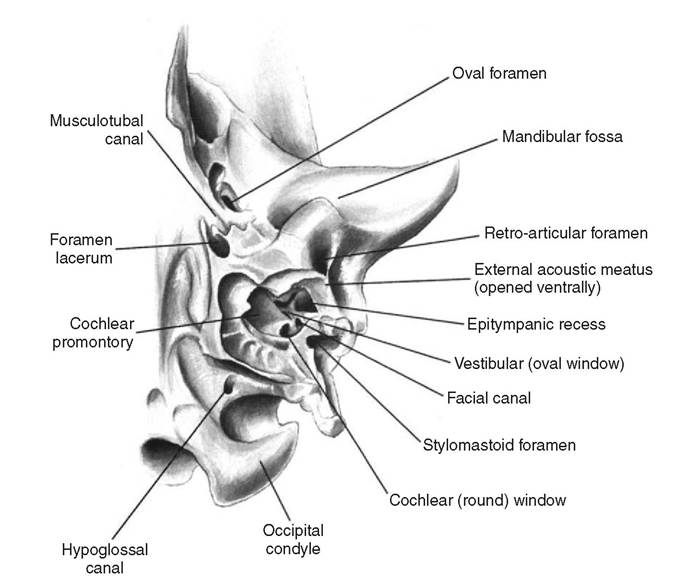
Figure 1-9
Internal anatomy of the left tympanic bulla, ventral view. The ventral wall of the bulla has been removed.
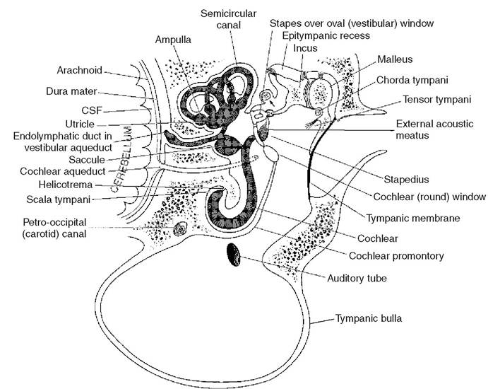
Figure 1-10
Schematic drawing of the internal and middle ear structures. The three ear ossicles form synovial joints with one another, supported by ligaments and two skeletal muscles.
Note the location of the facial nerve in relation to the middle ear cavity (arrow). Also shown are the relationships between subarachnoid space containing cerebrospinal fluid (CSF) and the inner ear via the cochlear aqueduct.An open slit facing the vestibular window is present in the bony facial canal (see Figures 1-9 and 1-10). This slit is the open part of the facial canal, which is covered by mucous membrane lining the tympanic bulla. The other fossae close to the external acoustic meatus accommodate the tensor tympani and stapedius muscles. The small middle ear bones—the malleus, incus, and stapes—are accommodated in an epitympanic recess located dorsal to the oval and vestibular window, immediately medial to the opening of the external acoustic meatus.
At the caudal aspect of the tympanic bulla is a large fissure called the tympanooccipital fissure, which is also called the petrobasilar or petro-occipital fissure. A medial foramen in the tympanooccipital fissure that leads into a canal between the occipital and temporal bones is called the caudal foramen lacerum. The bony canal that extends rostrally from the caudal foramen lacerum is called the carotid canal; it transmits the internal carotid artery and postganglionic sympathetic plexus from the superior cervical ganglion (also called the carotid plexus, made up of the carotid nerves; Figure 1-11). The carotid canal in its caudal extent also accommodates the ventral petrosal venous sinus (the caudal continuation of the cavernous venous sinus), which drains into the internal jugular and vertebral veins.
Ear Ossicles
The auditory ossicles—the malleus, incus, and stapes—are small movable bones that extend like a chain from the tympanic membrane and functionally connect the tympanic membrane with the vestibular (oval) window (see Figure 1-10). The ossicles consist of compact bone formed by endochondral ossification. They form synovial joints with each other.
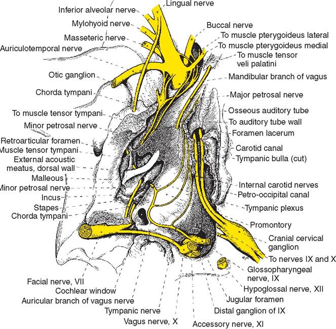
Figure 1-11
Nerves in relation to the middle ear cavity, ventral view, with most of the tympanic bulla removed.
(From Evans HE, ed: Miller’s Anatomy of the dog, ed 3, Philadelphia, 1993, WB Saunders.)The malleus is the most lateral bone. It consists of a head that articulates with the incus, a thin neck, and a long manubrium, or handle. The lateral aspect of the handle is concave and is embedded in the tympanic membrane, as described earlier.
Articulating with the medial part of the incus is the stapes. It is in direct contact with the perilymph fluid through its footplate (base of stapes) attachment in the oval window.
Vibrations of the tympanic membrane are transmitted through the chain of these auditory ossicles to the perilymph fluid within the vestibule. The vestibular window is approximately 18 to 20 times smaller in area than the tympanic membrane, contributing significantly to the amplification of sound waves by the ear ossicles.
The middle ear ossicles are associated with two very small skeletal muscles (see Figure 1-10). The tensor tympani muscle is a spherical muscle that attaches to the malleus by a short tendon. It is supplied by a branch of the trigeminal nerve. The stapedius, the smallest skeletal muscle in the body, is closely related to the facial nerve at its origin. This muscle is supplied by the facial nerve. Reflex contraction of these two muscles in response to loud noises results in fixation of the ear ossicles, damping vibrations. This protective reflex is called the tympanic reflex. It takes approximately 40 to 160 milliseconds for the tympanic reflex to occur.
More on the topic Middle Ear:
- Careful examination of a clean, dry ear canal in a dog or cat with otitis externa may reveal many conditions that affect the ear canal.
- Lateral Ear Canal Resection and Ear Canal Ablation
- in a recent book on the Slavonic peoples in the Middle East, the Russian Orientalist and historian Dmitrii E. Mishin examined a major but little- studied aspect of the historical relations between eastern Europe and the Middle East in Sakaliba (Slaviane) v islamskom mire v rannee srednevekove (Saqaliba:
- EAR DISORDERS
- Ear Cleaners
- The Normal Ear
- Structure and Functioning of Ear
- Stenotic Ear Canals
- Ear Therapy
- Ear Mites
- Hair in Ear Canals
- Structure of the Ear Canal
- Flushing of the Ear Canal
- Structure of the External Ear
- Vertical Ear Canal
- Inner Ear
- Neoplasia of the Ear Canals