Inner Ear
The main functions of the inner ear are receiving auditory signals and maintaining equilibrium. The inner ear is located within the osseous labyrinth of the petrous part of the temporal bone.
The membranous labyrinth consists of three primary parts: the cochlea, vestibule, and semicircular canals. The vestibule is divided into an utricle and a saccule. The vestibulocochlear nerve (cranial nerve VIII) supplies the membranous cochlea, vestibule, and semicircular canals (Figure 1-14).Osseous Anatomy
The petrosal part of the tympanic bone has an internal pyramid containing the internal acoustic meatus, the osseous and membranous labyrinths, and an external mastoid process. The pyramidal part of the petrous (meaning “rocklike”) temporal bone is the hardest bone in the body. The rostrodorsal surface of the pyramid is in contact with the cerebrum and hence is called the cerebral surface, whereas the caudomedial surface contacts the cerebellum and is called the cerebellar surface. The mid-part of the cerebellar surface of the pyramid exhibits a foramen called the internal acoustic meatus. The internal acoustic meatus in turn presents two tiny foramina: (1) a dorsally located foramen that leads into the osseous facial canal for the seventh cranial nerve and vestibular component of the eighth cranial nerve, and (2) a ventrally located foramen for the cochlear component of the eighth cranial nerve (see Figure 1-13). Because of the close association of the facial and eighth cranial nerves within the petrous temporal bone, the two nerves may be affected simultaneously by the same lesion.
The excavation within the petrous temporal bone, called the osseous labyrinth, is approximately 15 mm long. It is divided into three compartments: the cochlea, vestibule, and semicircular canals. The osseous vestibule is continuous with the subarachnoid space via the cochlear aqueduct (see Figure 1-10).
Via this duct, the perilymph is continuous with cerebrospinal fluid in the subarachnoid space surrounding the brain. Chronic middle ear infections can thus travel via the vestibular or cochlear window to affect the inner ear and eventually can spread via the cochlear aqueduct to the meninges. Within the osseous labyrinth, the membranous labyrinth is suspended in perilymph.Cochlea
The membranous labyrinth within the osseous labyrinth is an interconnecting system of epithelium-lined tubules and spaces filled with a clear fluid called endolymph. As
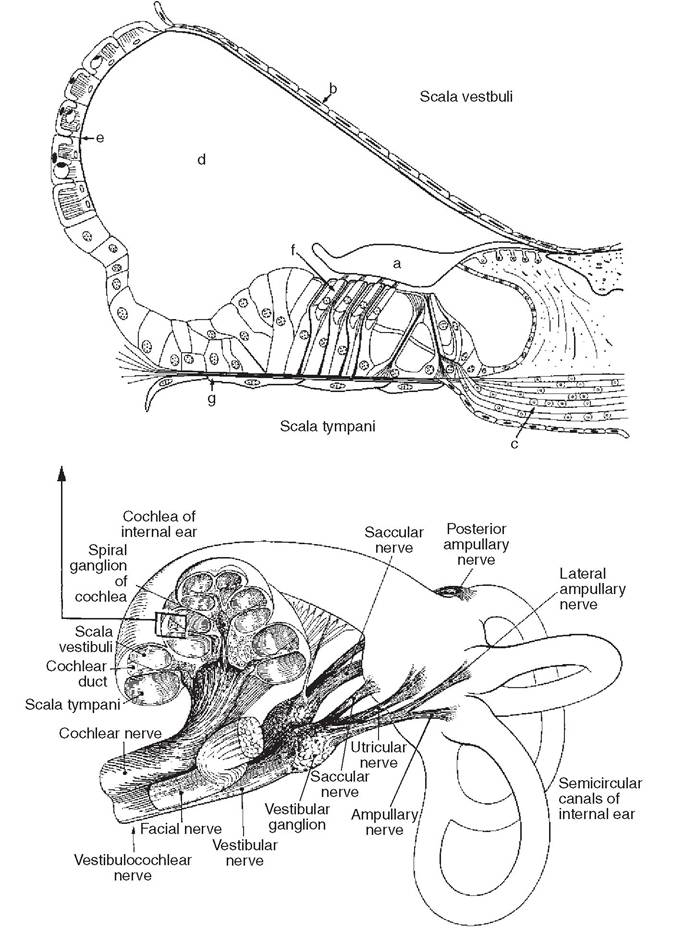
Figure 1-14
Schematic drawing of the inner ear. The membranous labyrinth is rotated 180 degrees ventrodorsally. Top panel shows the membranous cochlea in section and spiral organ (organ of Corti). a, Tectorial membrane; b, vestibular membrane; c, spiral ganglion of the cochlea; d, scala media; e, stria vascularis, which primarily produces endolymph; f, outer hair cells, which are sensory receptors for sound; g, basilar membrane. Lower panel shows innervation of the inner ear complex by branches of the eighth cranial nerve. mentioned before, the vestibule consists of the utricle and the saccule. These sacs communicate directly with each other and also with the semicircular and cochlear ducts. The cochlea is receptive to vibrations in endolymph, and the rest of the membranous labyrinth is associated with the function of equilibrium. Problems within the membranous labyrinth lead to signs of deafness and vestibular disease, including vestibular ataxia, circling, head tilting, strabismus, and nystagmus.
The bony cochlea winds around a hollow central axis (modiolus) in a dorsoven- tral direction. The modiolus accommodates the cochlear nerve (see Figure 1-14). The osseous or bony spiral lamina is a bony shelf that projects from the modiolus into the interior of the canal. Like the cochlea, it makes three and one quarter turns to end at the apex, or cupula (see Figure 1-13).
The bony spiral lamina reaches about halfway into the lumen of the bony cochlea, partially dividing its cavity into two parts, the dorsal scala vestibuli and the ventral scala tympani.The membranous cochlear duct is an epithelium-lined and endolymph-filled structure that extends from the osseous spiral lamina to the outer bony wall, completely dividing the scala vestibuli and scala tympani. The scala vestibuli and scala tympani are continuous with each other at the apex of the modiolus through a small foramen called the helicotrema. The cavity of the membranous cochlea, called the scala media, is filled by endolymph.
A specialized thickened epithelium in the basilar membrane constitutes the spiral organ (organ of Corti), in which cochlear nerve endings innervate the sensory hair cells. The hair cells are exposed to a specialized leaflike structure, the tectorial membrane. Vibrations of perilymph are transmitted to endolymph by the intervening (vestibular) membrane between the scala vestibuli and the scala media. The tectorial membrane in turn vibrates, touching the hair cells and initiating nerve impulses that are carried by the cochlear nerve to the brain.
Damage to the hair cells leads to hearing deficits or loss. Congenital deafness may be either perceptive (inner ear defect) or central (lesion of the auditory brain center). Acquired deafness may be either central or peripheral, resulting from chronic disease, normal aging, or drug use. Aminoglycoside therapy can lead to ototoxicity, a leading cause of iatrogenic hearing loss. White cats with congenital deafness have numerous cochlear abnormalities, including lesions in the spiral ganglion of the cochlear nerve.
Suggested Readings
Blauch B, Strafuss AC: Histologic relationships of the facial (7th) and vestibulocochlear (8th) cranial nerves within the petrous temporal bone in the dog, Am J Vet Res 35:481, 1974.
Evans HE: Miller’s Anatomy of the dog, ed 3, Philadelphia, 1993, WB Saunders.
Getty R: Sisson and Grossman’s The anatomy of the domestic animals, ed 5, vol 2, Philadelphia, 1975, WB Saunders.
King AS, Riley VA: A guide to the physiological and clinical anatomy of the head, ed 4, Liverpool, UK, 1980, University of Liverpool.
More on the topic Inner Ear:
- Careful examination of a clean, dry ear canal in a dog or cat with otitis externa may reveal many conditions that affect the ear canal.
- Lateral Ear Canal Resection and Ear Canal Ablation
- EAR DISORDERS
- The Normal Ear
- Stenotic Ear Canals
- Ear Therapy
- Ear Mites
- Ear Cleaners
- Hair in Ear Canals
- Structure of the Ear Canal
- Middle Ear
- Flushing of the Ear Canal
- Structure and Functioning of Ear
- Structure of the External Ear
- Vertical Ear Canal
- Neoplasia of the Ear Canals
- Examining the Ear Canal