Neoplasia of the Ear Canals
When a dog or cat with chronic otitis externa that does not respond to routine therapy is presented to the veterinarian, the clinician should be alert to the possibility of a tumor or growth in the ear canal.
Medical treatment for otitis externa may offer palliative relief, but the chronic recurrent nature of the disease is indicative of an underlying condition.When cytologic examination of otic exudates reveals large sheets of epithelial cells, neoplasia must be considered as a diagnosis. Chronic purulent otitis complicated by bacterial or yeast infection may also be obvious. Tumors occlude the ear canal and prevent drainage of exudates and therefore are often complicated by infection.
Symptomatology
Tumors arising in the external ear are often diagnosed only when they become large enough to be obvious. A tumor in the more distal portions of the ear canal may be obscured by wax (Figure 7-23), or the examiner may not look past the bend in the ear canal to see a tumor in the horizontal canal. Many tumors are extremely small, and in an ear canal with a very small diameter, they predispose to infection. The tumors
Figure 7-23
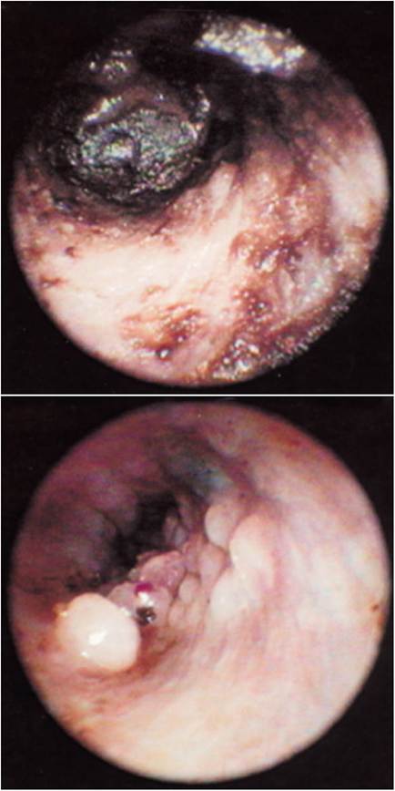
Many cerumen gland tumors are functional, producing wax in great quantities (top). The wax obscures the tumors until the ear is cleaned with a ceruminolytic (bottom).
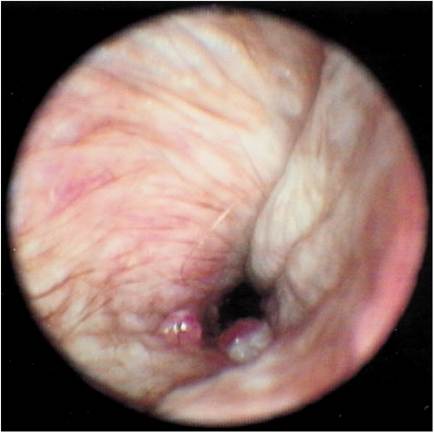
Figure 7-24
Ulcerated cerumen gland adenomas. This Golden Retriever had recurrent aural hematomas. The dog had been operated on three times previously for aural hematoma. When this dog was presented to the author for the fourth aural hematoma, a careful examination of the otherwise normal ear canal revealed these small ulcerated tumors.
Removal of these tumors at the time of aural hematoma surgery cured this dog of its head shaking.often have an ulcerated tip (Figure 7-24). Occlusion of the ear canal by a tumor can cause pain and discomfort. If the patient shakes the head excessively, aural hematoma may result. It is difficult to examine the horizontal canal for tumors without an instrument such as a video otoscope (Video Vetscope, MedRx, Inc., Largo, Florida), but if a tumor is found in the horizontal canal, it may explain the extreme discomfort the patient evidently experiences (Figure 7-25).
A tumor of the middle ear may present as vestibular disease with head tilt or nystagmus. If visualization is possible, as with a video otoscope, the tumor mass can be seen and samples taken for further diagnostic testing. Open-mouth rostrooccipi- tal radiographs or lateral views of the tympanic bullae (Figure 7-26) showing opacity of the osseous tympanic bulla may indicate a tumor or polyp of the middle ear. Osteolysis or fluid densities may also be seen. Computed tomography scans are also used to identify lesions within the middle ear.
Routine cleaning of the ear canal and careful examination of the skin surface of the ear canal may reveal small tumors. Good visualization and magnification are mandatory in identifying these very tiny early growths. Larger tumors may be confused with stenoses because they are large and occlude the lumen of the ear canal. Flat tumors may look like edematous ear canals, and sessile tumors have
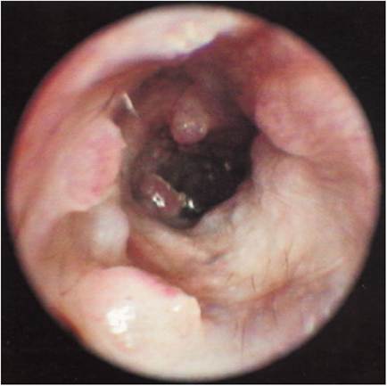
Figure 7-25
In addition to the tumors in the vertical ear canal of this Labrador Retriever, small cerumen gland tumors can be seen in the horizontal ear canal near the intact eardrum. These small tumors are hard to see with conventional otoscopy.
a cobblestone appearance (Figures 7-27 and 7-28). Some tumors are seen only when the otoscope is placed beyond a stenotic portion of the vertical ear canal.
Secondary infection is common in ears that harbor tumors. An obstructive tumor mass allows accumulation of cerumen and debris beyond it. Because the smooth, uniform epithelial surface has been permanently changed, accumulations of wax and serum are found in the deep recesses created by the uneven surface. These crevices promote bacteria and yeast growth. Continued antibiotic or anti-yeast treatment is warranted in the presence of tumors because of the secondary infections and inflammation commonly associated with aural neoplasms. If the eardrum is intact, flushing the ear canal with detergent ear cleaners is beneficial for removing the cerumen and debris. If the eardrum cannot be visualized, however, caution in selecting a non- ototoxic ear cleaner is warranted.
More on the topic Neoplasia of the Ear Canals:
- Hair in Ear Canals
- Assessing the Ear Canals
- Stenotic Ear Canals
- Careful examination of a clean, dry ear canal in a dog or cat with otitis externa may reveal many conditions that affect the ear canal.
- Lateral Ear Canal Resection and Ear Canal Ablation
- Neoplasia of the Pinna
- Mammary Neoplasia
- Pulmonary Neoplasia
- Hepatocellular Neoplasia
- Harderian Gland Neoplasia
- Neoplasia of the Reproductive System
- 38 Colorectal neoplasia in a dog
- Signature GEM Phenotypes: Molecular Pathology of Neoplasia
- Inner Ear
- Ear Mites
- Flushing of the Ear Canal
- One of the most common ailments of dogs seen in a veterinary practice is ear disease.