Gross Examination
On gross examination, tumors of the ear canal can have a variety of topographic features. They may be raised (Figure 7-29), pedunculated, broad based, lobulated, irregular (Figure 7-30), or ulcerated, or they may have a combination of these features.
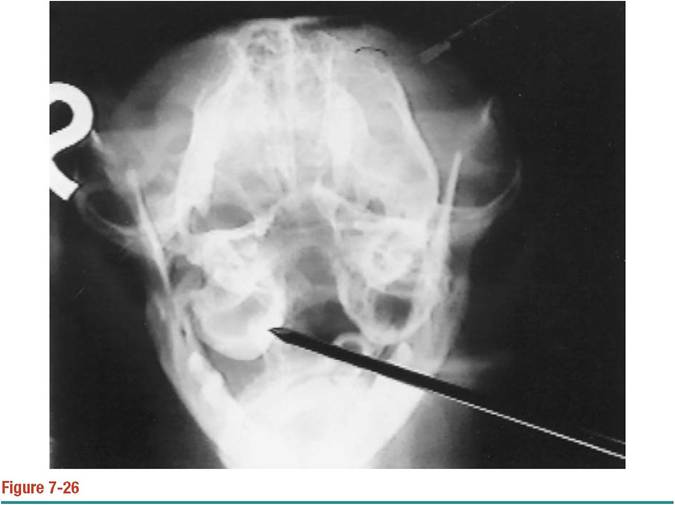
Open-mouth rostrooccipital radiograph of a cat with a unilateral nasopharyngeal polyp and otitis media.
Notice the thickened tympanic bulla and the opacified bulla lumen (pointer). (Courtesy Dr. Gary Lantz, Purdue University.)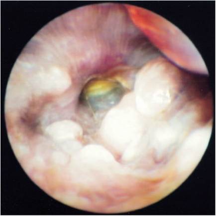
Figure 7-27
Flat, broad-based cerumen gland adenomas can be confused with edema or tissue maceration.
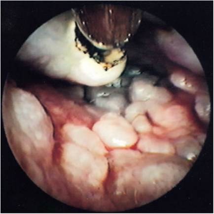
Figure 7-28
Protruding cerumen gland adenoma and cobblestone adenomas in the same ear canal as in Figure 7-27.
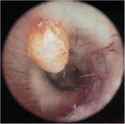
Figure 7-29
Solitary cerumen gland adenoma in an otherwise normal ear canal.
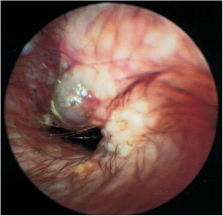
Figure 7-30
Irregular, broad-based cerumen gland adenoma. Smaller tumors surround the larger tumor mass.
For small tumors, tissue for histopathologic evaluation may be obtained by curetting the tumor with a sharp-edged curette (Figure 7-31). An endoscopic biopsy tool may be used to grasp and remove a section of tumor (Figure 7-32). In the case of a large solitary tumor or a diffuse spreading tumor mass, a portion of the ear canal is removed surgically, and the entire section of ear canal is submitted to the pathologist. Tissue architecture is preserved in this manner, and the pathologist can better characterize the lesion as benign or malignant.
Fortunately, the circumferential cartilage of the external ear canal provides a limiting barrier to the spread of most malignant tumors. If the ear tumor and the cartilage can be submitted to the pathologist, the invasiveness of the tumor can also be determined.Exfoliative cytologic evaluation is often adequate for differentiating inflammatory from hyperplastic and neoplastic lesions. A curette is used to obtain scrapings from a suspected tumor. If the cells indicate an inflammatory lesion (neutrophils, macrophages, lymphocytes, plasma cells, and eosinophils), appropriate antiinflammatory treatment may be instituted. If, however, clumps and clusters of large round cells are found, an epithelial neoplasm is a diagnostic possibility and the slide should be submitted to the pathologist for evaluation. Single spindle-shaped cells may indicate fibrosis or a nonepithelial type of neoplasm.
It has been theorized that chronic inflammation, more common in the dog than in the cat, may initiate progression of otic lesions from hyperplasia to dysplasia to neoplasia. It has also been speculated that bacterial degradation of the apocrine
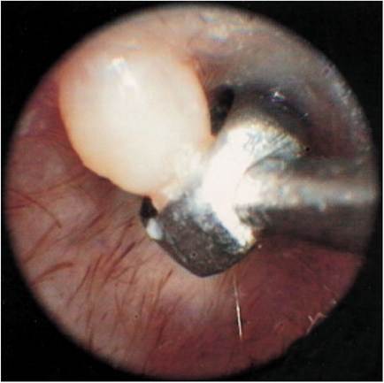
Figure 7-31
Removal of the tumor mass shown in Figure 7-29. A 6-mm dermal curette with a sharp edge has been placed over the mass. Angling the curette to cut the base of the tumor allows its removal.
secretions from the cerumen glands that become inspissated in the ear canal during episodes of otitis externa may result in increased carcinogenesis. Whether the presence of a tumor leads to the chronic inflammation or chronic inflammation leads to dysplastic changes and eventually to neoplastic changes is not known.
Ear canal tumors can develop in any of the skin structures lining the ear canal, including the squamous epithelium, ceruminous or sebaceous glands, and the mesenchymal structures (Figure 7-33). Tumors arising from the external ear canal and pinna are much more common than tumors arising from the middle or inner ear.
Much controversy exists concerning the incidence of the various tumors reported in dogs and cats. Because of the difficulty in obtaining samples from the ear canal, most samples submitted to diagnostic laboratories are submitted from university or referral surgeons and not from general practitioners. The number of tumors of the ear submitted for evaluation is therefore low.
Tumors of the ear canal are relatively uncommon in dogs and cats. In one report, of all tumors found in cats, only 1% to 2% were derived from aural tissue. In a second report, of all the canine aural tissue sent to different laboratories for histopathologic evaluation, 2% to 6% contained tumors. Generally, tumors of the ear canal in dogs tend to be benign, and tumors of the ear canal in the cat tend to be more malignant. Fortunately, malignant neoplasms of the ear canal rarely metastasize. Failure to remove a wide en bloc section of the ear canal, however, often results in
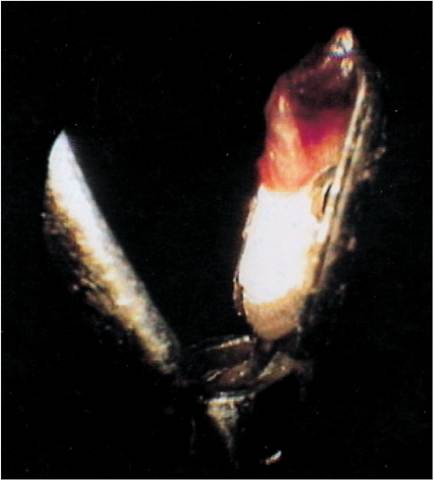
Figure 7-32
A 1.8-mm endoscopic biopsy tool was placed through the 2-mm working channel of the Video Vetscope to take a “bite” of the tumor for histopathologic evaluation.
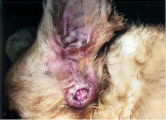
Figure 7-33
Tumor mass protruding from the horizontal canal of this Cocker Spaniel that had a previous vertical ear canal ablation. The mass was described by the pathologist as a liposarcoma.
local recurrence of tumor. Total ear canal ablation may be the best surgical solution to ear neoplasia.
Most reports indicate that benign cerumen gland adenoma is the most common tumor of the dog’s ear and that malignant ceruminous gland adenocarcinoma is the most common neoplasm of the cat’s ear. However, histopathologic differentiation of the two types of ceruminous neoplasms is not always clear. Other benign tumors found in the ear canal of dogs are polyps, papilloma, sebaceous gland adenoma, histiocytoma, plasmacytoma, benign melanoma, and fibroma.
Malignant canine neoplasms of the ear that have been reported are ceruminous gland adenocarcinomas, carcinoma of undetermined origin, squamous cell carcinoma, round cell tumor, sarcoma, malignant melanoma, and hemangiosarcoma.Benign tumors of the cat’s ear include polyps, cerumen gland adenoma, and papillomas. Malignant tumors of the cat’s ear are ceruminous gland adenocarcinoma, squamous cell carcinoma, carcinoma of undetermined origin, and sebaceous gland adenocarcinoma.
Tumors found in the middle ear are rare and include fibrosarcoma (Figure 7-34) and lymphoma.
Treatment of neoplasia found in the ear canals of dogs and cats requires surgical intervention so that the tumor mass can be completely excised. Newer modalities such as electrosurgery and laser surgery through a video otoscope (Video Vetscope, MedRx, Inc., Largo, Florida) make removal of ear canal tumors easier.
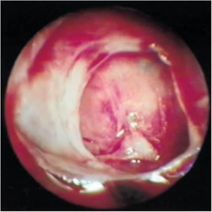
Figure 7-34
Middle ear fibrosarcoma. A fleshy mass was seen in the middle ear. Myringotomy revealed a large infiltrating mass filling the tympanic bulla. Histopathologic analysis of a biopsy specimen revealed fibrosarcoma.
Histopathologic evaluation of removed tissues helps in providing a prognosis of the disease. For malignancies, follow-up radiation therapy has been shown to be effective in preventing recurrence of many ear canal tumors, especially cerumen gland adenocarcinoma of cats.
Suggested Readings
Gotthelf LN: Secondary otitis media: an often overlooked condition, Canine Pract 20:14-20, 1995.
Logas D, Rosychuck RAW, Merchant SR: Diseases of the ear canal, Vet Clin North Am Small Anim Pract 24(5):905-980, 1994.
London CA, Dubilzeig RR, Vail DM, et al: Evaluation of dogs and cats with tumors of the ear canal: 145 cases (1978-1992), J Am Vet Med Assoc 208:1413-1418, 1996.
Moisan PG, Watson GL: Ceruminous gland tumors in dogs and cats: a review of 124 cases, J Am Anim Hosp Assoc 32:449-453, 1996.
Rogers KS: Tumors of the ear canal, Vet Clin North Am Small Anim Pract 18:859-868, 1988.
Theon AP, Barthez PY, Madewell BR, et al: Radiation therapy of ceruminous gland carcinomas in dogs and cats, J Am Vet Med Assoc 205:566-569, 1994.
van der Gaag I: The pathology of the external ear canal in dogs and cats, Vet Q 8:307-317, 1986.
More on the topic Gross Examination:
- Elite-formation: the Army, the Lineage and the Examination System
- 49 Gestational T rophoblastic Disease
- Tympanic Bulla
- Diagnosis of Bovine Tuberculosis in Zambia
- 32 Colonic vascular ectasia in a dog
- Pneumocystis murina Infection: Pneumocystosis
- INFORMING INTERVIEW
- TECHNICAL FACTORS OF NEEDLE ELECTROMYOGRAPHY
- REFERENCES
- Judicial reasoning and contextual imperatives