Tympanic Bulla
The osteum of the auditory tube lies in a rostrodorsomedial location in the tympanic bulla, coursing anteriorly. The auditory tube is lined by pseudostratified ciliated columnar epithelium containing goblet cells.
The auditory tube opens into the nasopharynx and equalizes air pressure on either side of the tympanic membrane. On the medial wall of the tympanic cavity is the promontory, a bony shelf that houses the bony labyrinth (see Figure 1-10).The central space of the labyrinth, called the vestibule, is continuous with the subarachnoid space via the cochlear aqueduct (see Figure 1-10). It is filled with perilymph, which is similar to cerebrospinal fluid. The vestibule connects to the three semicircular canals and the cochlea.
On the lateral side of the promontory is a thin bony plate containing two windows or fenestrations. The oval or vestibular window lies on the dorsolateral surface of the promontory immediately adjacent to the pars flaccida. It attaches to the base of the stapes and connects to the vestibule. Vibrations are transmitted to the endolymph contained within the cochlea from the tympanic membrane via the ear ossicle chain to the oval window. The round window, an opening in the bony shelf of the osseous cochlea, is covered by a secondary tympanic membrane. It is found in the anterior end of the vestibule, just ventral to the smaller oval window. This membrane permits vibrations that have already passed to sensory receptors to be dissipated into the airfilled tympanic bulla.
The middle ear cavity of the cat is divided by a septum into two separate tympanic cavities (Figure 1-12). In the small dorsolateral compartment lie the auditory ossicles, the osteum of the auditory tube, and the eardrum. The larger ventromedial compartment is an air-filled tympanic bulla. The two compartments of the tympanic
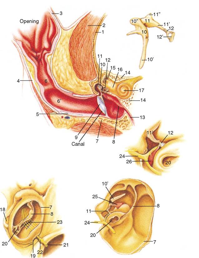
Figure 1-12
Schematic views of the cat middle and internal ear. (From Hudson LC, Hamilton WP: Atlas of feline anatomy for veterinarians, Philadelphia, 1993, WB Saunders.)
bulla communicate via a small passage located dorsally near the cochlear window. This intervening bony septum should be perforated when necessary for proper drainage of the middle ear cavity. Rough handling of the bony septum may result in damage to the postganglionic sympathetic nerves. The nerves, which are visible submucosally as fine strands over the cochlear promontory, should be avoided during surgical removal of the septum in the cat.
More on the topic Tympanic Bulla:
- Safe Drugs for the Tympanic Bulla
- Imaging of the Tympanic Bulla
- Flushing and Suctioning the Bulla
- Bulla Infusion and Topical Therapy
- Middle Ear
- Assessing the Eardrum
- Otitis Media Prevents Healing
- Treatment
- Causes of Rupture
- Otitis Media
- Gross Examination
- Myringotomy
- Is the Eardrum Ruptured?
- Common ear ailments of dogs and cats can be seen in up to 50% of patients in areas of humid climates during some or all of the patient’s life.
- Canine Polyps
- Lateral Ear Canal Resection and Ear Canal Ablation
- Treatment of Otitis Media
- Conventional Radiography
- Secondary Otitis Media in Dogs