Canine Polyps
The dog only rarely develops a nasopharyngeal polyp originating from the mucosa of the tympanic bulla or from the eustachian tube. There have been reports of polyps occluding the airway as they grow through the eustachian tube into the nasopharynx.
In one report, a canine nasopharyngeal polyp was so large that an incision into the soft palate was required to access it at the external opening of the auditory tube in order to grasp and remove it.9 Other reports demonstrate that there are nasopharyngeal polyps that protrude through the eardrum into the ear canal in the same manner as the feline inflammatory polyp. Canine inflammatory polyps have been reported to occur bilaterally on occasion. One report indicated polyps only in older male dogs.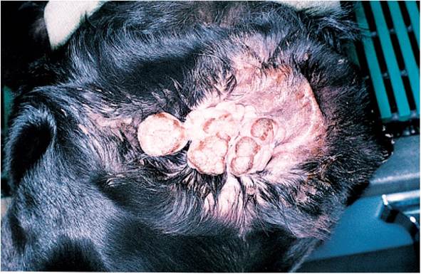
Figure 16-8
Canine inflammatory polyp. This Labrador Retriever was presented with both pinnae extending laterally from his head. Inflammatory polyps were present in both ears. The white polyp mass visible in this photo filled the external opening to the ear canal; as it grew, the polyp molded to the epithelial folds of the tragus of the pinna. The polyps were easily separated from the pinna. Each had a thick vascular stalk that extended through the entire ear canal to the middle ear. The polyps were easily removed by excision, and the stalks were removed using traction.
The significance of this is unclear.10 The author has seen several canine aural polyps that grow very large, and they grow to the outside of the ear canal, often molding to the epithelial folds at the tragus (Figure 16-8).
Traction removal has been successful in canine polyps seen by the author, perhaps because they can be grasped with stronger forceps and can be visualized easier than feline polyps (Figures 16-9 and 16-10).
Surgical treatment of canine polyps requires total ear canal ablation and lateral bulla osteotomy (TECA-LBO) rather than ventral bulla osteotomy, as in cats, because of the chronic nature of the external ear canal pathology that is found in most dogs with inflammatory polyps.Most dog polyps are not true polyps but huge ceruminous gland adenomas. This type of tumor often has a broad base originating from the epithelial surface of the external ear canal. The eardrum is usually intact in these cases. The rapid growth and the position of this tumor mass in the ear canal make the diagnosis confusing. Pinch biopsies of these masses are sometimes reported as polyps, because there is an inflammatory stroma with inflammatory cells covered by epithelium. When the entire mass is submitted for histopathology, the dilated ceruminous glands become apparent.
This type of otic mass cannot be removed using traction because of its broad attachment. Lateral ear canal resection with excision from the ear canal at the base of the tumor is the preferred method of removing large ceruminous gland adenomas (Figure 16-11).
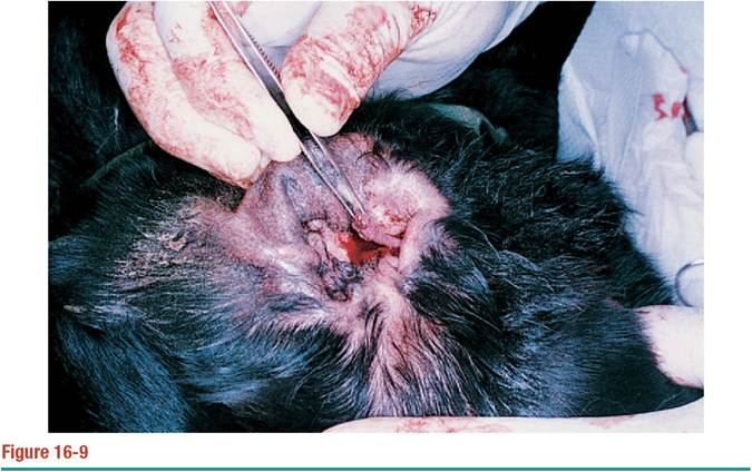
When the polyp is removed by excision, the peduncle remains (grasped in thumb forceps). Unless this vascular supply is removed at the time of surgery, the polyp is likely to grow back.
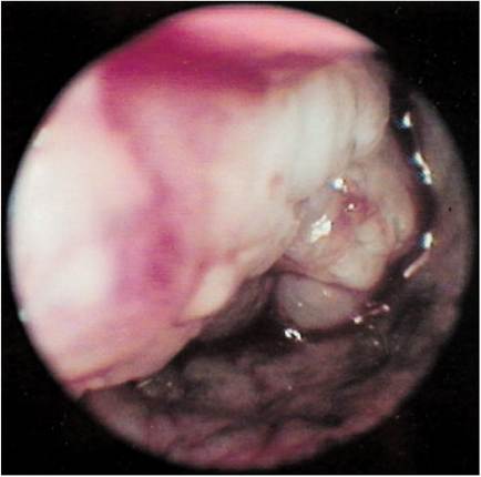
Figure 16-10
Endoscopic view of the stalk that remained in Figure 16-9. The long stalk, or peduncle, in this polyp extended from the middle ear mucosa through the external ear canal.
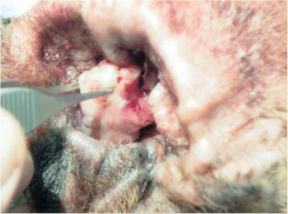
Figure 16-11
Cerumen gland adenoma in the vertical canal of a dog. This type of tumor is often confused with a true nasopharyngeal polyp. The difference is that these tumors are attached by a broad base to the ear canal epithelium.
The eardrum in many of these dogs is normal, and there is no otitis media.References
1. Allen HS, Broussard J, Noone K: Nasopharyngeal diseases in cats: a retrospective study of 53 cases (1991-1998), J Am Anim Hosp Assoc 35(6):457-461, 1999.
2. Faulkner JE, Budsberg SC: Results of ventral bulla osteotomy for treatment of ear polyps in cats, J Am Anim Hosp Assoc 26(5):496-499, 1990.
3. Trevor PB, Martin RA: Tympanic bulla osteotomy for the treatment of middle ear disease in cats: 19 cases (1984-1991), J Am Vet Med Assoc 202(1):123-128, 1993.
4. Veir JK, Lappin MR, Foley JE, et al: Feline inflammatory polyps: historical, clinical, and PCR findings for feline calicivirus and feline herpesvirus-1 in 28 cases, J Feline Med Surg 4(4):195-199, 2002.
5. Boothe HW: Surgery of the tympanic bulla (otitis media and nasopharyngeal polyps), Prob Vet Med 3(2):254-269, 1991.
6. Stanley BJ: Management of nasopharyngeal polyps in cats, Proceedings Waltham Feline Medicine Symposium, 1998.
7. Mortellaro CM, Alfieri C, DeFrancesco I, et al: Perendoscopic trans-tympanic excision (PTTE) of ear canal polyps in cats: 10 case reports, World Small Animal Veterinary Association Proceedings, 2001.
8. Anderson DM, White RAS, Robinson RK: Management of inflammatory polyps in 37 cats, Vet Rec 147(24):684-687, 2000.
9. Fingland RB, Gratzek A, Vorhies MW, et al: Nasopharyngeal polyp in a dog, J Am Anim Hosp Assoc 29:311-314, 1993.
10. Pratschke KM: Inflammatory polyps of the middle ear in 5 dogs, Vet Surg 32(3):292-296, 2003.
More on the topic Canine Polyps:
- Anorectal Polyps
- Feline Polyps
- CANINE DISTEMPER IN CARNIVORES
- canine Parvoviral enteritis
- INFECTIOUS CANINE HEPATITIS
- 16 Inflammatory Polyps
- Canine inflammatory bowel disease activity index (CIBDAI)
- Paul Bloom, DVM, DACVD, DAB VP (Canine an d Fel ine Specialty)
- Signalment and History
- Index
- Microbiology
- Tumors of the Ear Canal
- Atopic Dermatitis
- Otitis media is a common disease process that often goes unrecognized in most veterinary practices.
- CONTENTS
- Abnormal Radiographic Findings
- Rhinosinusitis
- Treatment
- Summary
- Diagnostic Tests