Is the Eardrum Ruptured?
Several techniques have been described to determine the integrity of the TM when it cannot be visualized in an ear with a stenotic external ear canal.4 A small-diameter (31/2 to 5 Fr) catheter can be inserted into the ear canal until it stops.
It is then extended and retracted to get a feel for the rigidity of the “stop.” If there is a spongy feel, the eardrum is intact. If there is a definite hard feel to the “stop,” the eardrum is ruptured and the catheter is hitting the medial wall of the tympanic bulla. This technique should be practiced on cadaver specimens to acquire the necessary sensitivity.Tympanometry uses a sensor that measures the compliance of the eardrum in response to sound waves. It is not practical to perform this test in the veterinary clinic, however, because it is still a research tool in animals.
An easy, indirect method for determining the integrity of the eardrum is to infuse warmed, very dilute povidone-iodine solution (or dilute fluorescein solution) into the ear canal with the anesthetized dog or cat in lateral recumbency. If the orange or yellow-green flushing fluid comes out of the nose or if the patient snorts out this solution through the oropharynx when pressure is applied by the flushing fluid, the eardrum is ruptured (Figure 14-8). The fluid has flowed from the external ear canal through the ruptured eardrum, into the tympanic bulla, and through the auditory tube into the nasopharynx.
Another technique used by some is to fill the ear canal of a patient in lateral recumbency with the suspected ruptured eardrum up with warmed saline and to insert the tip of the video otscope into the ear canal. By looking through the clear
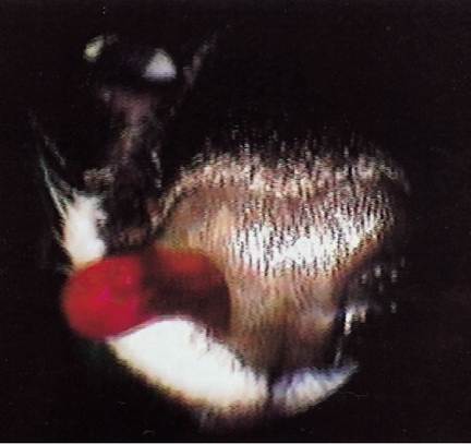
Figure 14-8
Nasal drainage of dilute povidone iodine flushed from the middle ear during ear cleaning. fluid, if air bubbles rise from the ear canal while the animal breathes, the eardrum is ruptured.
Air from the nasopharynx rises through the auditory tube into the tympanic bulla to escape from the middle ear through a ruptured eardrum.Positive contrast canalography has been described as a method for detecting a ruptured TM in dogs with otitis media.5 Two to 5 ml of dilute iodinated contrast agent is instilled into the ear canals of these anesthetized patients while in lateral recumbency with the affected ear up. The author uses 0.3 ml of Hypaque 50% or similar contrast agent in 2.7 ml of saline. In a stenotic ear canal, a 31/2- or 5-Fr catheter is threaded into the stenosis if possible. Contrast agent is then infused beyond the stenosis. An open-mouth view of the bullae is then taken, using a horizontal x-ray beam. If the eardrum is intact, there will be a distinct contrast/air interface at the eardrum. If the eardrum is not intact, the contrast material will enter the bulla, and a continuous column of contrast will extend into the bulla.
In a study of this technique, eardrums of cadaver dogs with normal ear canals and intact eardrums were intentionally ruptured and contrast material was introduced into the external ear canal. In every case, contrast media entered the tympanic bulla and was detected by a radiograph. In clinical otitis media cases, positive-contrast canalography was positive in most of the cases where the eardrum was determined to be ruptured otoscopically, and it was positive for other cases in which the eardrum appeared to be intact otoscopically. In normal ears, canalography was more accurate for detecting iatrogenic TM perforation than otoscopy.5
More on the topic Is the Eardrum Ruptured?:
- Assessing the Eardrum
- Veterinarians are often faced with the problem of ruptured eardrums.
- Healing Process
- Evaluation of the Patient
- Secondary Otitis Media in Dogs
- Signalment and History
- Failure of Epithelial Migration
- Ear Therapy
- Myringotomy
- Appendix Ear Product Formulary
- Topical Ototoxicity