Myringotomy
To diagnose and treat patients with otitis media, it is sometimes necessary to perform a myringotomy to get a cytology specimen and allow for culture and antibiotic sensitivity testing on the material trapped behind the eardrum.
If there is fluid pressure pushing on the eardrum or negative pressure retracting it, perforation of the eardrum using a controlled myringotomy incision immediately relieves the intense pain associated with these pressure changes.To perform a myringotomy, the patient is anesthetized and the external ear canal is thoroughly cleaned with a disinfectant such as dilute povidone iodine. The ear canal is then dried using suction. A sterile, rigid polypropylene catheter is cut to a 60-degree angle with a surgery blade to provide a sharp point. A long spinal needle can also be used to puncture the eardrum. The tip of the cut catheter is advanced under good visualization, and the pars tensa is punctured at either the 5 or 7 o’clock position in order to remain away from the germinal epithelium and blood vessels overlying the manubrium of the malleus (Figure 14-10).
Alternatively, a small Buck curette (2 mm) can be used to make a hole in the eardrum. This instrument makes a larger hole in the eardrum and is more difficult to direct accurately to the proper site for puncture. This technique may be used to create a large hole in the eardrum to allow middle ear exudates to drain into the horizontal canal and to prevent pressure gradients from recurring. Larger instruments should not be used for myringotomy because they cause tearing of the eardrum.
Many veterinary practices are using CO2 lasers to make the myringotomy incision. A 0.8 mm ? 180 mm rigid tip or a long, flexible Teflon tip can be inserted through the working channel of the video otoscope and can be advanced to the eardrum. Applying a pulsed, low wattage (3 or 4 watts) laser impulse melts the eardrum.
The advantage of laser myringotomy is that the tip does not have to touch the eardrum, so there is less chance of contamination of the bulla with external ear canal material.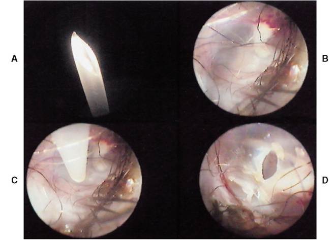
Figure 14-10
Myringotomy. A, A 5-Fr polypropylene catheter cut to a 45-degree angle with a scalpel blade. B, Surgical landmarks are identified. Blood vessels identify the pars flaccida and the hooked bone is the malleus. These structures should be avoided. C, The sharp pointed catheter is advanced along the floor of the horizontal canal with the bevel up. In the dog the tympanic membrane is at a 45-degree angle to the horizontal canal. D, Advancing the catheter and pushing it through the eardrum creates a “smiler” type of incision.
In addition, the hole made by the laser is circular and takes longer to heal, which is sometimes beneficial in providing drainage (Figure 14-11).
Fluid under pressure may freely flow into the horizontal canal as the perforation begins, and it should be suctioned to ensure that the myringotomy incision is large enough to accommodate a 3½- or 5-Fr catheter. In the case of suppurative otitis media, myringotomy serves to decrease the fluid pressure behind the eardrum. The fluid escapes into the external ear canal and may continue to drain for several days, so during therapy the ear canals need to be flushed to remove this debris. The catheter is advanced through the incised TM and directed ventrally into the bulla; gentle suction is then used to retrieve any material within the bulla. If a spinal needle was used, the stylet is withdrawn before suctioning. If the bulla is dry, 1 or 2 cc of normal saline can be infused into the bulla and then immediately retrieved. This material is submitted for cytology, bacterial culture, and antibiotic sensitivity.
Certain antibiotics or corticosteroids can be infused directly into the tympanic bulla through the myringotomy incision, providing high topical levels within the tympanic bulla.
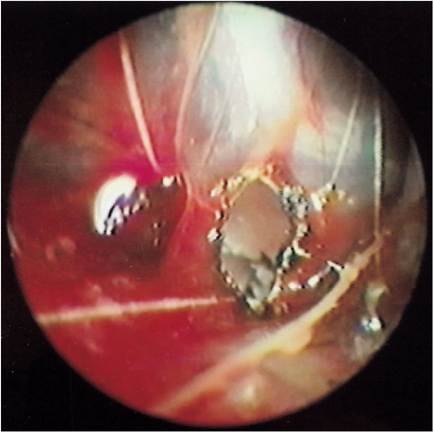
Figure 14-11
Laser myringotomy creates a more circular hole because the eardrum is vaporized. Laser myringotomy heals slower than catheter myringotomy, which may be an advantage for repeated treatments.
More on the topic Myringotomy:
- Myringotomy
- Myringotomy
- Sample Collection
- Causes of Rupture
- Epithelial Migration
- Treatment of Otitis Media
- Tympanic Bulla
- Cytological Evaluation
- Is the Eardrum Ruptured?
- Magnetic Resonance Imaging (MRI)