Trauma
Trauma from any external cause can affect the ears. Whether due to fight wounds, trauma to the pinna from being hit by a car, or surgical trauma, a tissue reaction triggered by a traumatic injury may lead to problems in the ear canals.
Wounds
Underlying the ear canal epithelium is a subcutaneous layer of dermis and a cylindrical to conical layer of elastic cartilage surrounded by muscles, blood vessels, and nerves. Trauma to any one of these tissues causes intense inflammation and bleeding, and may lead to acute stenosis of the ear canal from the edema that follows inflammation. In most cases the stenosis resolves when the tissue in the damaged portion of the ear canal heals. In other cases, especially in traumatic injury to the cartilage, the healing may be very slow and return of normal anatomic architecture incomplete. The stenotic canal that remains is the result of contracture of fibrous connective tissue or deformation of cartilage during healing. An example of the deformity of cartilage can be seen in an untreated aural hematoma in which the shape of the pinnal cartilage has been permanently changed. When the cartilaginous cylindrical shape has been compromised, the lumen of the ear canal may be obliterated. Fistulous tracts may appear around the site of facial trauma that may communicate with the ear canal (Figures 7-17 and 7-18).
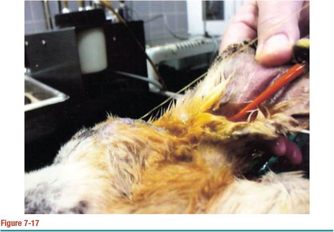
Traumatic facial wound of a Beagle resulted in a communication from the facial wound to the ear canal. A red rubber catheter has been placed in the ear canal and dilute povidone-iodine is being flushed into the ear canal. As the ear canal is being flushed, the flush solution exits from the facial wound in a forceful stream parallel to the red rubber catheter.
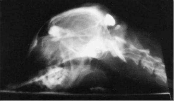
Figure 7-18
Radiograph showing infusion of iodinated contrast medium into the facial wound shown in Figure 7-17.
Contrast material can be seen filling the facial wound site, the ear canal, the tympanic bulla, and the subcutaneous region in the occipital area.Surgical trauma from ear canal surgery can also lead to postoperative complications. Procedures such as total ear canal ablation often result in fistulous tracts emanating from the incompletely removed lining of the middle ear with unresolved infection. Vertical ear canal ablation or the Zepp lateral ear resection surgery requires cutting through cartilage. Suturing through cartilage incites an inflammatory reaction. Hyaluronic acid, which is released into the surrounding tissue from the cut cartilaginous matrix, stimulates intense inflammation and swelling; therefore, suturing skin to skin over the cartilage is advised.
Trauma induced by blunt force injury to the head and neck can result in a ruptured eardrum (Figure 7-19). Fresh bleeding from the ear may be seen on examination. The ear canals in such cases must be examined for the presence of an eardrum and accumulation of blood behind the eardrum.
Trauma from Cotton-Tipped Applicators
Cotton-tipped applicators (Q-tips) are often used for removal of ear wax in humans. The dog’s ear canal is long, bent, and tapered, so the tip of an ear swab inserted into the vertical canal quickly approaches the diameter of the ear canal as it is advanced.
Trauma from the cotton-tipped applicator occurs as the ear canal is plugged by the cotton tip. The ear canal is “scraped” by the abrasive cellulose or synthetic fiber.
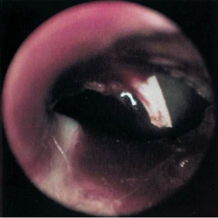
Figure 7-19
The patient, a mixed terrier, was hit by a car and had blood coming out of the ear. Examination showed that the patient had a ruptured eardrum. The manubrium of the malleus is prominently displayed.
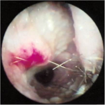
Figure 7-20
A cotton-tipped applicator used in this ear resulted in denuding the epithelial surface with the abrasive material. Acid-type ear cleaners will cause severe pain when they contact the ulcer.
In an infected ear, the epithelium is edematous and very often friable, so that only mild pressure from a cotton-tipped applicator can result in ulceration (Figure 7-20). When the ear canal becomes ulcerated, microorganisms can flourish as growthenhancing nutrients, such as blood components and serum proteins, are released into the ear canal.
Cotton-tipped applicators also tend to push any material that may be ahead of the cotton tip farther down the ear canal, packing cerumen into the horizontal canal. If enough pressure is applied, this material may be pushed right through the eardrum. Cotton-tipped applicators themselves may perforate the eardrum if they are pushed too quickly into an ear, creating a traumatic myringotomy (Figure 7-21).
Cotton-tipped applicators are available in a variety of tip sizes. Veterinarians should use only the small applicators in the ears of small animals. Obtaining rollsmear cytologic samples (see Chapter 2) for determining the organisms present in otic exudates can be accomplished by inserting the small cotton-tipped applicator into the horizontal ear canal through an otoscope cone and pulling the swab slowly along the ear canal toward the pinna.
Use of cotton-tipped applicators should be restricted to removal of liquids. The absorptive capacity of the cotton tip allows liquid to be absorbed. Simply inserting the tip very carefully into an ear filled with liquid (e.g., flush solutions, pus, mucus) while restricting movement of the applicator enables liquids to be absorbed into the tip. Repeated insertions of applicators are required until a tip is dry when removed.
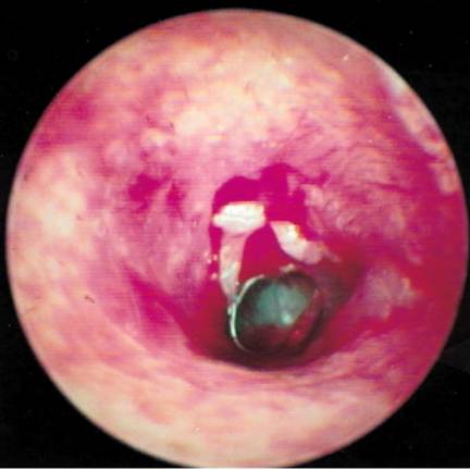
Figure 7-21
This cat's ear was filled with wax and debris associated with ear mites. The material was flushed out, but when a cotton-tipped applicator was put into the ear, it popped the eardrum.
Trauma from Instrumentation
Many instruments are used in the ear canal by veterinarians, groomers, and pet owners.
Curettes are used to remove wax and tenacious material from the epidermal layer. Various rigid catheters are put into the ear canals for flushing and suctioning debris; sharp edges on cut urethral or feeding tubes will cut the epithelium of the ear canal. Curved hemostats are used for pulling hair from the ear canals; hemostats tend to crush tissue and, when improperly used, may cause damage to the ear canal. Water pumps, such as the Water Pik, provide a forceful stream of ear cleaning solution; increased water pressure in the ear canal can rupture an already macerated, friable eardrum.Because most procedures done in the ear canal are performed without good visibility, trauma invariably occurs. Instruments and catheters are fairly small, and some bleeding and discomfort may be evident after the procedure. Various medications and ear cleaners may burn the ear tissue when applied. Most traumatic injuries from instruments heal rapidly. The exception is perforation of the eardrum (Figure 7-22).
When traumatic perforation of the eardrum occurs, it is intensely painful. The patient exhibits clinical signs and pain that were not previously present. The amount of exudates in the ear canal may rapidly increase, and the otitis externa being treated appears worse. Traumatic myringotomy heals in approximately 2 to 3 weeks, if iatrogenic otitis media has not been created. In the presence of an otitis externa, however, pushing exudates into the middle ear predisposes to otitis media.
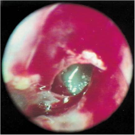
Figure 7-22
Ulceration and iatrogenic perforation of the eardrum in a Poodle's ear. This large hole in the eardrum may never completely heal. Medications that may be potentially ototoxic should not be used in this ear.
More on the topic Trauma:
- CHILDHOOD TRAUMA
- Pinnal Trauma
- Head Trauma
- Concept of trauma
- Trauma
- Nonaccidental Trauma
- Literature and Art, Nostalgia and Trauma
- Quantifying Violence: Assessing the Prevalence of Trauma
- Diaphragmatic disorders are usually congenital, though acquireddefects due to direct trauma or neurological injury (diaphragmatic palsy) are not uncommon.
- In mental life nothing which had once been formed can perish - that everything is somehow preserved and that in suitable circumstances... it can once more be brought to light... on condition that the organ of the mind has remained intact and that its tissues have not been damaged by trauma or inflammation.
- Symmetrical Pinnal Alopecia
- Violence and the Archaeological Record
- Later Stone Age Violence in Context
- 22.7 DIABETES MELLITU
- Acute Spinal Cord Dysfunction
- Physical Injury