The Normal Ear
To become familiar with the appearance of the normal ear canal and eardrum, the veterinarian should perform a thorough ear examination on every patient anesthetized for any reason (such as ovariohysterectomy or a dental procedure).
It is necessary to know (1) what the normal ear canal should look like, (2) where the eardrum should be located, if it is not readily visible, and (3) how to distinguish normal cerumen from otic exudates in order to determine whether the ear is affected by disease.Ear Canal
The normal ear canal epithelium should be light pink with small superficial blood vessels visible (Figure 2-1). Small amounts of cerumen coating the epithelium give the surface a glistening appearance. Cerumen is a normal part of a healthy ear and should not be regarded as pathological unless it is excessive. Hairs are seen along the canal, being more numerous in the vertical canal (Figure 2-2).
The dog’s ear canal gently bends approximately 75 degrees as it changes from the vertical to the horizontal (Figure 2-3). With the dog in the standing position, the examiner should gently place traction ventrally on the pinna; the ear canal will straighten out, because the normal underlying cartilage is soft and pliable. The otoscope cone should be advanced into the horizontal canal as the canal straightens. This technique enables the horizontal canal to be examined. In the anesthetized patient in lateral recumbency, the examiner can straighten out the canal by lifting the pinna vertically to the point of elevating the entire head.
Hairs are almost absent from the horizontal canal in most dogs. However, a clump of long, bristly hairs is often found at the distal end of the horizontal canal and may cause discomfort (Figure 2-4). An accumulation of wax may be seen along the
Figure 2-1
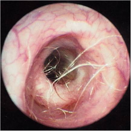
Normal ear canal of the dog.
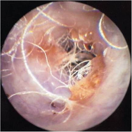
Figure 2-2
Numerous hairs and wax found in the vertical canal of a poodle.
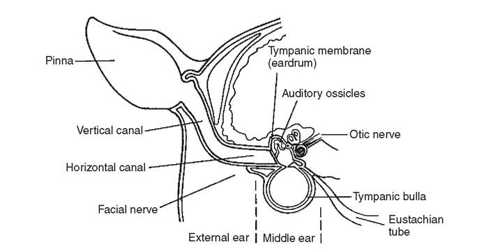
Figure 2-3
The anatomy of the dog's ear canal. The vertical canal bends at approximately 75 degrees to become the horizontal canal.
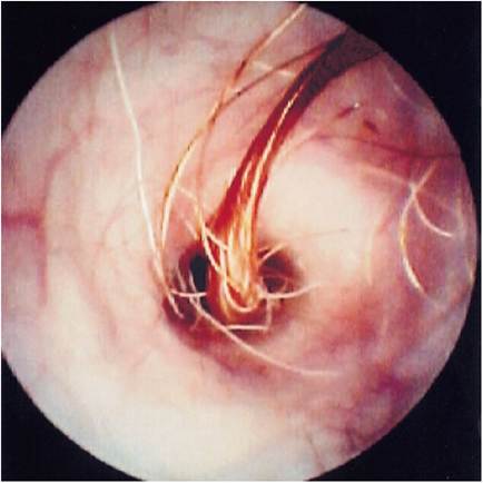
Figure 2-4
A tuft of long, thick, bristly hairs is sometimes found in the horizontal canal originating near the annulus of the eardrum. These hairs can be irritating and may become the matrix for wax plugs.
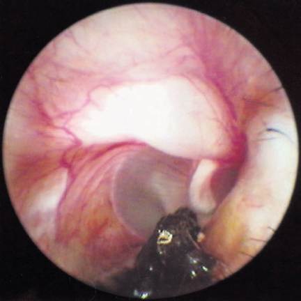
Figure 2-5
Accumulation of wax along the ventral floor of the horizontal canal. This wax is helpful for orienting the dorsoventral axis when examining the ear canals.
ventral floor of the horizontal canal (Figure 2-5). As the otoscope is advanced in the horizontal canal, the tympanic membrane, if present, should become visible.
Tympanic Membrane
The normal tympanic membrane in both the dog and the cat appears as a thin, transparent to translucent, reflective end of the horizontal ear canal (Figures 2-6 and 2-7). The normal eardrum has been described as having a pale, “rice paper” appearance. The outline of the footplate (manubrium) of the malleus is seen attached to the medial side of the tympanic membrane, frequently bulging outward (pars tensa). The malleus is seen as a thin rectangular white bone originating from the dorsal portion of the tympanic membrane and extending ventrally halfway across the membrane. The malleus is oriented dorsoventrally. The free distal end of the manubrium may have a gentle curve or a “hook” that points rostrally. This feature aids in distinguishing the right ear from the left ear on photographs of the eardrum.
Examination of the dorsal portion of the tympanic membrane (pars flaccida) reveals an opaque, pink or white, loose membrane often containing a network of small blood vessels that extend across the tympanic membrane (Figure 2-8). This “vascular strip” often has an edematous appearance, and this “pretympanic bleb” may obstruct visualization of dorsal portions of the eardrum. In many cases of otitis media, the vascular strip is destroyed, removing from the germinal epithelium of the
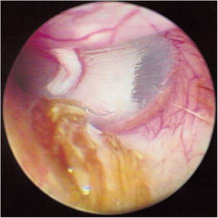
Figure 2-6
Normal canine left eardrum. The yellow wax is in the ventral portion of the horizontal canal. The footplate of the malleus can be clearly seen through the rice paper-thin pars tensa. The malleus has a “hook” that points rostrally. In the dorsal portion of the eardrum is the pars flaccida with the blood vessels that supply the epithelium over the malleus.
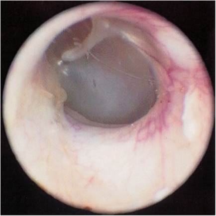
Figure 2-7
Normal feline eardrum.
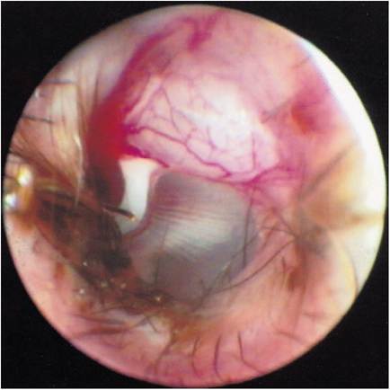
Figure 2-8
Prominent pars flaccida forming the “vascular strip.” This provides the blood supply to the eardrum. Radial collagen fibers can be seen in the pars tensa.
eardrum the blood supply that is required for the eardrum to heal. If the vascular strip is unaffected by disease or trauma, the damaged eardrum has a much better chance of completely healing with time.
The tympanic membrane in dogs is oriented at an acute angle of as much as 45 degrees to the long axis of the horizontal canal, making visualization of the entire eardrum difficult.
Often, the lower portion of the eardrum’s attachment to the horizontal canal is obscured from view due to a depression of the floor of the horizontal canal, forming a fornix. Some dogs may have negative pressure in the tympanic bulla, which causes retraction of the eardrum into the bulla and makes examination difficult. A condition known as a “false middle ear” has been described, in which the tympanic membrane is actually stretched inward into the bulla from exudates or negative pressure and becomes attached to the lining of the middle ear. In this condition, the entire middle ear cavity is obliterated. When such an ear is examined with the otoscope, no eardrum is visible, but a deep, dark hole can be seen.
In the anesthetized patient in lateral recumbency, the pinna is lifted vertically to straighten the curved ear canal, which makes advancing of the otoscope cone easier. The tip of the instrument can be advanced closer to the eardrum. The cat’s tympanic membrane is easily visualized because the eardrum is oriented in a plane forming a 90-degree angle with the short ear canal.
More on the topic The Normal Ear:
- Careful examination of a clean, dry ear canal in a dog or cat with otitis externa may reveal many conditions that affect the ear canal.
- Lateral Ear Canal Resection and Ear Canal Ablation
- Normal Cytology
- CARE OF NORMAL NEWBORN
- Ear Cleaners
- Working Toward a New “Normal”
- NORMAL GAIT IS CYCLICAL AND SYMMETRIC
- NORMAL ACID-BASE REGULATION
- EAR DISORDERS
- Stenotic Ear Canals
- Ear Therapy
- Normal Development
- Normal Nutritio
- Ear Mites
- 9 Gastrointestinal physiology - the normal stomach and small intestines
- Technique and Normal Findings