Normal Cytology
Cytology is not performed to identify the disorder otitis externa; this diagnosis is based on history and clinical signs. Instead, cytology is used to characterize the nature of otic exudate in order to identify potential primary causes and clinically significant perpetuating factors.
Thus, cytology is rarely performed on normal asymptomatic ear canals. However, since determining the relative significance of cytologic findings requires a thorough understanding of what is and is not normally found in the external ear canals of dogs and cats, a discussion of normal cytology is appropriate.The epithelial lining of the normal ear canal is coated with a thin layer of cerumen, the combined product of secretions by modified apocrine glands (ceruminous glands) and sebaceous glands. Cerumen forms a protective barrier, which traps debris, organisms, hair, and desquamated corneocytes. A sample taken from a normal ear canal should yield minimal material consisting mostly of waxy, yellow cerumen, exfoliated epithelium, resident microorganisms, and little else.
Because cerumen is predominantly lipid, the sample does not take up much stain; on gross examination a stained slide from a normal ear should be nearly colorless. Normal cornified squamous epithelial cells are seen on microscopic examination as sheets of lightly stained, basophilic keratin. The cells may roll up on themselves during smear preparation, resulting in a deeper staining, shardlike appearance. Desquamated keratinocytes may also contain melanin granules, which appear as tiny yellow to brown ovoid or round structures. Recognition of these melanin granules as a normal finding is important; otherwise, the structures may easily be misidentified as cocci or small rods colonizing the surface of the keratinocyte. Melanin granules do not take up stain; therefore, granules can be differentiated from purple-stained bacteria by focusing up and down through the cell until the true color of the structure is recognized.
The external ear canals of dogs and cats contain small numbers of normal resident bacteria. Coagulase-negative Staphylococcus spp., coagulase-positive Staphylococcus spp., and Streptococcus spp. are the most frequently isolated bacteria from normal ear canals. With the exception of Corynebacterium, rod-shaped bacteria are rarely found in normal ear canals.4,6,11 Any bacteria found in the presence of leukocytes should be considered abnormal.5-7 Because stain precipitate can resemble coccoid bacteria, high-oil magnification is necessary to visualize morphologic characteristics of bacteria. Clinically significant organisms should be symmetrical, have a distinct smooth edge, and be uniformly stained; they are typically present in pairs or chains of bacteria equal in size (Figure 3-3). In contrast, debris and precipitate vary in size and may be asymmetrical, irregular, and granular in appearance.
Another common finding on otic cytology is basophilic staining yeast, ranging in size from 2.0 μm ? 4.0 μm up to 6.0 μm ? 7.0 μm.12 For comparison, canine red blood cells are approximately 7.0 μm in diameter; feline red blood cells are 5 μm. The most commonly encountered yeast exhibits unipolar budding, which creates the commonly described “peanut,” “snowman,” or “footprint” shape, easily recognizable as Malassezia (Figure 3-4). Although these organisms are normal residents of the canine and feline ear canal, under the appropriate circumstances Malassezia can
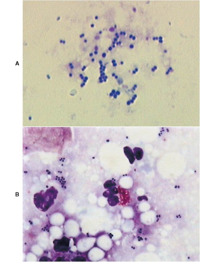
Figure 3-3
A, Cluster of paired coccoid bacteria photographed under high-oil immersion lens (100? objective). B, Paired coccoid bacteria with neutrophils and an eosinophil (400? objective).
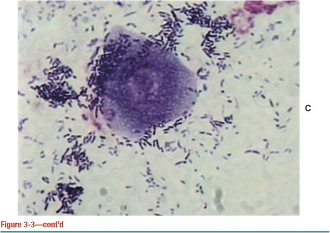
C, Numerous rods and a keratinized epithelial cell (1000? objective).
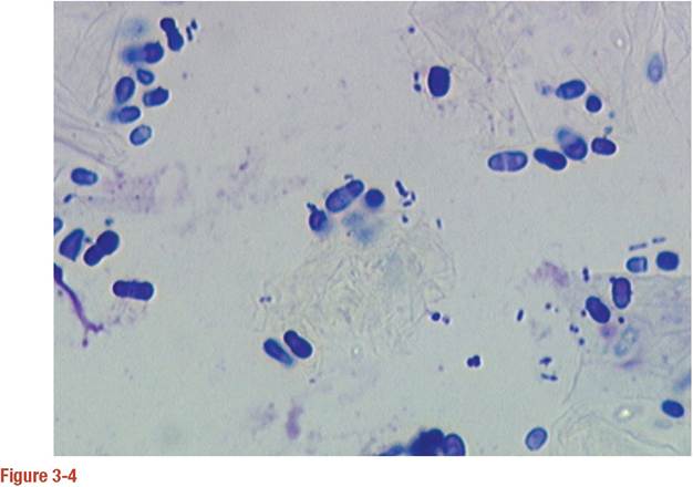
Malassezia pachydermatis with occasional paired coccoid bacteria in background. Note the oval shape of nonbudding yeast and unipolar budding of actively replicating organisms.
become important opportunistic pathogens, contributing directly to the severity of clinical signs, as well as to the progression and perpetuation of disease.
Because bacteria and yeast are considered normal inhabitants of the external ear canal, veterinarians need to determine whether the bacteria present cytologically are clinically relevant. In contrast, any finding of leukocytes on cytology is considered abnormal. The finding of bacteria that have been engulfed by phagocytic neutrophils and macrophages is one clear indication of relevance (Figure 3-5). The absence of leukocytes does not rule out the role of bacteria or yeast as pathogens. In general, heavy colonization of the canal by opportunistic pathogens is reflected in the number of bacteria seen per high-powered field. This semiquantitative method for determining clinical significance is discussed in detail in the sections on abnormal cytology.
More on the topic Normal Cytology:
- Abnormal Cytology
- Cytology
- Cytology and Histopatnology of the Ear in Health and Disease
- The Normal Ear
- CARE OF NORMAL NEWBORN
- Working Toward a New “Normal”
- NORMAL GAIT IS CYCLICAL AND SYMMETRIC
- NORMAL ACID-BASE REGULATION
- Normal Development
- Normal Nutritio
- 9 Gastrointestinal physiology - the normal stomach and small intestines
- Technique and Normal Findings
- chapter 1 Mindstyle='font-variant:normal !important;text-transform:uppercase'>
- CHAPTER 6 α1Antitrypsin Therapy Increases CD4+ Lymphocytes to Normal Values in HIV-1 Patients
- Roman Law in the Plang=EN-US style='font-variant:normal !important; text-transform:uppercase'>rovinces