MYOPATHIES
Myopathies are generally hereditary, though clinical manifestations may be delayed for variable period of time. Important myopathies are listed in Table 18.41. It
TABLE 18.41: Common myopathies in children
• HereditaryZcongenital
- Muscular dystrophies
Duchhene muscular dystrophy (DMD/BMD)
- Others: LGMD, FSHD, myotonic MD, congenital MD
- Developmental: Congenital myopathies, benign hypotonia
- Metabolic: Carnitine deficiency, GSDs
- Mitochondrial myopathies, e.g.
MELAS, MERRF• Acquired
- Endocrinal: Hypothyroidism, steroid-induced
- Metabolic: Hypokalemic periodic paralysis
- Inhammatory: Dermatomyositis
LGMD: Limb girdle muscular dystrophy, FSHD: Facioscapulohumeral dystrophy, GSD: Glycogen storage disorders, MELAS: Myoclonic epilepsy, lactic acidosis and recurrent stroke-like syndromes, MERRF: Mitochondrial encephalopathy with Red-Ragged fibers
is essential to understand difference between common 'muscular dystrophies' and rare 'congenital myopathies', as both may present at any age.
Muscular dystrophies are essentially: (a) hereditary, (b) progressive, and (c) primary myopathies of normally formed muscles, excluding secondary involvement in neuropathies.
Congenital myopathies are: (a) hereditary or non- hereditary, (b) non-progressive, and (c) developmental defect or maturational arrest in formation of normal muscle.
Duchenne muscular dystrophy (DMD) is the commonest (1:3600 male live births) hereditary myopathy in Indian children, characterized by progressive weakness of mainly proximal lower limb muscles with pseudohypertrophy, leading to severe motor disability, loss of ambulation and usually death in second decade of life. A milder variant, i.e. Becker muscular dystrophy (BMD) presents at a later age with more gradual course. DMD and BMD together are also termed as Dystrophinopathies.
Etiology: DMD/BMD is an X-linked recessive disease with affected males and carrier females, due to deletion, duplication of point mutation of one or more exons of the DMD gene, located on Xp21 locus (DMD gene is the longest gene in human with 79 exons). However, 30% cases may be new mutations where mother is not a carrier. Girls are very rarely affected, if having Turner syndrome (single X chromosome) or random inactivation of normal chromosome (Lyon hypothesis).
Primary product of DMD gene is an important cytoskeletal protein - Dystrophin, present in skeletal and smooth muscles, brain and retina. This dystrophin is absent or severely deficient in DMD, while those with BMD have milder deficiency.
Clinical manifestations: Boys are asymptomatic at birth with normal early development including walking. In a typical case of DMD, weakness usually begins at ~ 2-3 years of age with:
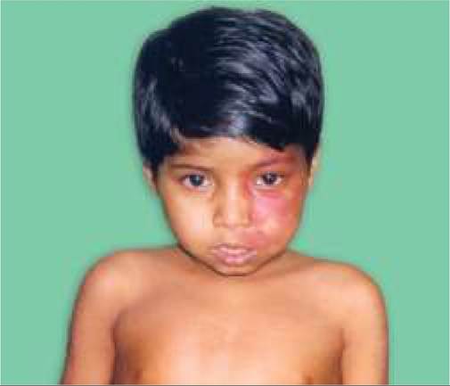
Fig. 18.21A and B: Duchenne muscular dystrophy: Psuedohypertrophy of calf muscles.
• Mild waddling (Trendelenburg) gait with a lordotic posture to adopt for gluteal weakness.
• Positive Gower sign, i.e. difficulty in standing from recumbent position due to pelvic girdle weakness. When asked to do so, he typically turns to his side, lifts his trunk up by supporting his weight on his arms and then stands up as if climbing upon his body with hand support.
• Pseudohypertrophy of calf muscles with wasting of thigh muscles (Fig. 18.21). Tongue and forearms may also develop pseudohypertrophy. Distal deep jerks, e.g. ankle and brachioradialis are stronger than proximal knee or biceps/ticeps jerks.
• Progressive weakness of proximal muscles including respiratory involvement with weak cough and recurrent respiratory infections, and/or pharyngeal muscle weakness with recurrent aspirations, nasal twang and regurgitation. Distal muscle functions, e.g. writing, etc. are spared till terminal stage.
• Associated features, e.g. cardiomyopathy and mild mental retardation or learning disabilities are present in nearly all cases with variable severity, not necessarily correlating with severity of muscular disease. Cardiomyopathy usually appears at ~10 years, though CCF and arrhythmia are uncommon till terminal stage and usually precipitated by intercurrent infections. Intermittent gastric dilatation with abdominal pain and vomiting is common.
Course: Patient gradually becomes bedridden by 7-10 years of age with development of contractures and scoliosis and die by 15-20 years due to respiratory failure, aspiration or CCF.
Carriers are asymptomatic but may have myalgia, cramps and mild calf hypertrophy in 10% cases.
Becker muscular dystrophy, the milder variant, usually present in late childhood with inability to raise the hand above shoulder (e.g. during combing the hair), remain ambulatory till 15-20 years and survive till 4th-5th decade.
Diagnosis may be made clinically on the basis of: (a) positive Gower sign, (b) proximal muscle weakness with pseudohypertrophy, and (c) similar family history, supported by markedly elevated CPK levels. CPK levels are high (gt;10 times of normal) even in asymptomatic cases and assist in early diagnosis of the disease in siblings. Carriers have mild CPK elevation.
Confirmation requires genetic diagnosis with multiplex PCR or MPLA to detect deletion or duplication in the DMD gene, though next generation exome sequencing (NGS) may be needed to detect point mutations in some cases.
Muscle biopsy is no longer necessary in genetically proven cases, but may reveal: ( a) scattered degenerating and regenerating myofibrils, (b) endomysial connective tissue proliferation, and (c) focal mononuclear infiltration. It also helps to differentiate DMD (no dystrophin staining) from Becker dystrophy (reduced and patchy staining) on immunohistochemistry.
Dystrophin levels are lt;3% of normal in classic DMD and 20-90% in Becker dystrophy.
EMG is indicated only in doubtful cases with near normal CPK levels, which reveals low amplitude and duration of motor action potential.
Carrier detection is possible by: (a) pedigree charting, (b) mild elevation of CPK levels in 80% cases, (c) muscle biopsy in suspected carriers, and (d) detection of dystrophin mutations in blood samples.
Prenatal diagnosis is possible as early as by 12 weeks PCR/MPLA testing on chorionic villi samples.
Management is entirely supportive at present, though experimental studies after discovery of dystrophin molecule are promising.
• Supportive treatment includes:
- Nutritional support (but avoid obesity),
- Physiotherapy to prevent/delay contractures,
- Ambulatory support (wheel chair /orthotic braces),
- Calcium supplements to prevent osteoporosis,
- Treatment of respiratory infections and CCF,
- Psychological support.
• Steroids (PO prednisolone 0.75 mg/kg/d for 10 days a month) have shown temporary improvement in muscle weakness for 4-6 months by decelerating the rate of myofiber necrosis. However, it does not alter the ultimate course and exact role is controversial.
• Newer and promising interventions in selected cases include:
- Antisense oligonucleotide drugs, e.g. Drisapersen and eteplirsen, which induce exon-skipping during splicing of mRNA to produce a shorter but potentially functional dystrophin protein and have been approved for use in selected cases.
- Ataluren, another drug approved for non-sense mutation, binds to ribosomes and enables stop codon read through.
- Other experimental therapies include: (a) myoblast transfer therapy from a near relative, or (b) recombinant dystrophin gene therapy.
• Prenatal screening and counseling is the best strategy so far to counter this disease.
Some other important myopathies in children are as follows:
Limb girdle muscular dystrophy (LGMD) is a group of slowly progressive inherited disorders, mainly affecting muscles of hip and shoulder girdle.
Some of them are autosomal dominant (LGMD-1) while others are recessive (LGMD-2). A mutant dystrophin-related protein (sarcoglycan) has been associated with autosomal recessive disease with characteristic sarcoglycanopathy on muscle biopsy.Most cases present in adolescence with gluteal muscle weakness leading to back pain and lordosis and progress gradually to non-ambulatory state by 3rd decade. Calf hypertrophy is common. Simultaneous weakness of shoulder and neck muscles is common, though facial, ocular and bulbar paralysis is rare. Intelligence is normal. Facioscapulohumeral dystrophy (autosomal dominant involving chromosome 4q35) is a relatively benign disorder, which manifests at around puberty with facial weakness (inability to close eyes, smile, whistle), shoulder girdle weakness (scapular winging) and upper limb weakness (inability to comb the hair). Weakness is slowly progressive with gradual involvement of pelvic girdle (Gower sign).
Unlike DMD, pseudohypertrophy is absent, cardiomyopathy is rare and intelligence is spared. CPK is mildly elevated and EMG/muscle biopsy suggests coexisting neurogenic pathology. Diagnosis rests on genetic testing and treatment is supportive.
Myotonic muscular dystrophy (autosomal dominant involving DMKP gene on chromosome 19) is the second commonest muscular dystrophy after DMD in Europe, USA and Australia, though rare in India.
Clinically, these cases manifest with—(a) typical facial features (tented upper lip), (b) progressive weakness of distal muscles, e.g. hands, starting after infancy. Presence of myotonia, i.e. slow relaxation of muscles after contraction (e.g. slow release of grip) is pathognomonic though develops only after 10 years of age. Multi-systemic involvement with mental retardation, cardiomyopathy, cataracts, endocrinal disorders and immunodeficiency is common.
A rare variant presents in neonatal period with severe hypotonia (floppy infant), facial wasting and feeding/ breathing problems.
Most cases die in infancy.CPK and EMG are usually normal in children, while EMG may demonstrate myotonia in older patients. Diagnosis rests on genetic testing and treatment is supportive.
Congenital muscular dystrophies (autosomal recessive) encompass many distinct disorders, all with severe diffuse hypotonia (floppy infant) and proximal muscle weakness at birth. Facial, ocular and bulbar muscles are relatively spared. However, further course is slowly progressive and many cases are able to stand and walk with support in late childhood, unless contractures have developed. CNS involvement is common with mental retardation and white matter changes on MRI. CPK is moderated elevated and muscle biopsy shows signs of muscular dystrophy with merosin deficiency on immunohistochemistry.
Fukuyama disease, the commonest congenital muscular dystrophy, is the second commonest cause of muscular dystrophy in Japan (after DMD), associated with severe cardiomyopathy, microcephaly, mental retardation, seizures and ocular abnormalities.
Developmental muscle disorders may be isolated or associated with other congenital anomalies. Some important developmental disorders include:
Congenital absence of muscles is usually unilateral, e.g. absence of pectoralis major (Poland syndrome with associated amastia, rib deformities, webbed fingers and radial nerve aplasia), sternocleidomastoid (torticollis), depressor angularis oris (facial asymmetry on crying), etc. Asymptomatic absence of palmaris longus muscle of ventral forearm is present in ~ 30% population.
Proteus syndrome (Elephant man) is an extremely rare disorder of ecto/mesodermal overgrowth, with
asymmetric overgrowth of extremities including muscle hypertrophy, bony thickening, angiomas, cutaneous lesions, and hemi-megalencephaly.
Benign congenital hypotonia is clinical term to denote generalized non-progressive hypotonia without weakness, hyporeflexia or developmental delay. Intelligence is normal. Diagnosis rests on exclusion of congenital myopathies on muscle biopsy. Prognosis is good, except persistence of hypotonia which may lead to recurrent joint dislocations or injuries. Many of them are circus artists or gymnasts.
Congenital myopathies is a heterogeneous group of rare disorders, all characterized by defective development or arrested maturation of muscles (d/d muscle dystrophies, in which initial muscle development is normal). Important examples are myotubular myopathy (X-linked), congenital muscle fiber-type disproportion (usually sporadic), nemaline- rod myopathy (AR), central core disease (AD).
Clinically, congenital myopathies are non-progressive and usually present—(a) at birth or in early infancy as floppy child, or (b) in late childhood, with delayed motor development or defective ambulation (proximal girdle weakness). Facial and ocular muscles are frequently affected with feeding and breathing difficulties in severe cases. Unlike DMD, affected muscles are wasted rather than pseudohypertrophic. Central core disease is consistently associated with malignant hyperthermia.
Diagnosis is based on muscle biopsy including electron microscopy, as CPK levels are frequently normal and EMG changes are non-specific.
Treatment is non-specific and supportive. Severely affected children die in early infancy due to aspiration or respiratory insufficiency. Mild cases are non-progressive but may require orthotic/ambulation support.
Inflammatory myopathies, characterized by skeletal muscle inflammation gt; muscle fiber damage gt;weakness; may be postinfectious (influenza or HIV virus, toxoplasmosis, bacterial) or autoimmune in origin (dermatomyositis, polymyositis, eosinophilic myositis). Dermatomyositis is the leading cause of inflammatory myopathy in children.
Endocrinal myopathies in children are not as common as in adults, seen in hypothyroidism, thyrotoxicosis, primary hyperparathyroidism and steroid-excess, e.g. prolonged steroid therapy or Cushing disease. Most of them are associated with muscle wasting, except Kocher Debre semelaigne syndrome, i.e. generalized pseudohypertrophy in some cases of hypothyroidism.
Metabolic myopathies may be seen in many inborn errors of metabolism, e.g. glycogenoses (type I-V and VII) or lipid disorders (Carnitine deficiency), discussed elsewhere. Two other important causes include:
Hypokalemic periodic paralysis, an autosomal dominant disease, characterized by recurrent episodes of sudden and generalized weakness of skeletal muscles, with transient hypokalemia. Each episode lasts for few minutes to few hours and patient is normal between attacks.
Serum potassium, EMG and muscle biopsy (vacuolar myopathy) is abnormal only during attacks. Attacks may become more frequent in adulthood, leading to permanent and progressive myopathy. Hypokalemia due to other causes, e.g. renal tubular acidosis or hyperaldosteronism (Conn syndrome) may also cause transient and intermittent paralysis.
Rarely, similar episodes are known with transient hyperkalemia (hyperkalemic periodic paralysis), specially in cases of chronic renal failure, or excessive potassium supplementation.
Mitochondrial myopathies are part of systemic mitochondrial disorders due to point mutations in mitochondrial or nuclear DNA, leading to altered aerobic metabolism in various cells with secondary histopathological abnormalities. Brain and muscles are most affected. Mitochondrial DNA mutations disorders are exclusively inherited from mothers. Important mitochondrial myopathies include:
• MELAS (Mitochondrial myopathy, Encephalopathy, Lactic Acidosis and Stroke-like episodes) usually presents as a degenerative disorder with—(a) delayed motor and cognitive development, (b) recurrent hemiplegia/hemianopia (stroke-like episodes) and/ or seizures, (c) episodic vomiting, seizures and progressive dementia. Muscle biopsy reveals typical ragged-red fibers. Most cases manifest in late childhood and die in few years.
• MERRF (Mitochondrial Encephalopathy with RedRagged Fibers) present with myoclonic seizures, progressive myopathy (with red-ragged fibers on biopsy) and cerebellar signs in late childhood or adults with/without dementia and other features.
18.19
More on the topic MYOPATHIES:
- Myopathies
- Dystrophic Myopathies
- TECHNICAL FACTORS OF NEEDLE ELECTROMYOGRAPHY
- Physical Examination
- Electrodiagnostic Evaluation of the Floppy Infant
- Degenerative Changes in the Nervous System
- Disorders of the Endocrine System
- Spontaneous Hemorrhagic Necrosis of the Central Nervous System of Fetal Hamsters
- Microscopic Polyangiitis
- Infantile Botulism