Pulmonary Management
Pulmonary complications are recognized as the leading cause of mortality in childhood neuromuscular disease. Respiratory insufficiency in neuromuscular disease results from a number of factors, including: respiratory muscle weakness and fatigue, alteration of respiratory system mechanics, and impairment of a central control of respiration.
Progressive muscle weakness and fatigue lead to restrictive lung disease and ultimately to hypoventilation, hypercarbia, and respiratory failure. Increased inelastic load on respiratory muscles occurs because of chest wall stiffness, airway secretions, and ineffective cough mechanism. This may result in atelectasis and increased airway resistance, and kyphoscoliosis can further alter respiratory mechanics. Defects in central control of respiration may be secondary to hypoxemia and hypercarbia, associated with severe restrictive lung disease. Significant nocturnal decreases in partial pressure of oxygen, as well as elevations in arterial partial pressure of carbon dioxide, occur in more severe restrictive lung disease. Hypercapnia or hypoxemia occurring at night may have a role in reducing daytime central respiratory drive. A chronic increase in the bicarbonate pool may blunt the stimulus to breathe, generated by respiratory acidosis and perpetuating the hypercapnic state. Expiratory muscle weakness may produce ineffective cough, problems with clearance of secretions, and predisposition to pulmonary infections.Respiratory failure may present acutely or insidiously. Respiratory difficulties in the delivery room or early infancy may be seen in acute infantile type I SMA, myotubular myopathy, congenital hypomyelinating neuropathy, congenital infantile myasthenia, congenital myotonic muscular dystrophy, transitory neonatal myasthenia, and severe neurogenic arthrogryposis. In most other childhood neuromuscular diseases, the respiratory insufficiency develops more insidiously unless an acute decompensation occurs from an event such as an aspiration episode or acute onset of weakness, as seen in Guillain-Barre syndrome, botulism, and myasthenic syndromes.
Signs and symptoms of significant respiratory difficulties may include subcostal retractions, accessory respiratory muscle recruitment, nasal flaring, exertional dyspnea or dyspnea at rest, orthopnea, generalized fatigue, and paradoxic breathing patterns. A history of nightmares, morning headaches, and daytime drowsiness may indicate nocturnal hypoventilation with sleep-disordered breathing. Pulmonary function tests have been used to help in the decision-making process regarding the institution of mechanical ventilation. In a study of 53 patients with proximal myopathy, hypercapnia occurred when the maximal inspiratory pressure was less than 30% of predicted and when vital capacity was less than 55% of predicted (141). Other authors (142,143) have noted lower values for vital capacity measurements in their patients with DMD at the time they require institution of mechanical ventilatory support. Hahn and colleagues (144) have reported the predicted value of maximal static airway pressures in predicting impending respiratory failure. Splaingard (145) reviewed a series of 40 patients with a diverse group of neuromuscular disease conditions. They noted that all their patients who required mechanical ventilation had a vital capacity of ≤25%, with at least one of the following associated findings: PaCO2 >than 55 mmHg, recurrent atelectasis or pneumonia, moderate dyspnea at rest, or congestive heart failure.Noninvasive forms of both positive and negative pressure ventilation are being increasingly applied to children with neuromuscular diseases. Initially, patients may require ventilatory support for only part of the day. Noninvasive nocturnal ventilation has become a widely accepted clinical practice, providing ventilatory assistance for patients while sleeping and allowing them to breathe on their own during the day. Intermittent ventilation may ameliorate symptoms of respiratory failure, reduce hypercarbia, increase oxygenation (even during periods off the ventilator), and prolong survival in patients with neuromuscular disease.
The long-term use of noninvasive ventilation (Fig. 12.19) may be associated with fewer complications than ventilation via a tracheostomy; however, bulbar muscle function should be adequate for safe swallowing (117). Ventilatory support has allowed prolonged survival and acceptable quality of life in SMA I, SMA II, and DMD (143,146,147,148).Improved pulmonary toilet and clearance of secretions can be achieved with assisted cough; deep breathing; and setup spirometry, percussion, and postural
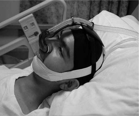
Figure 12.19 Noninvasive ventilatory support using bilevel positive airway pressure and nasal pillows mask interface in young adult with Duchenne muscular dystrophy.
drainage, and, in more severe cases, the additional use of interpulmonary percussive ventilation (IPV), given two to three times daily.
More on the topic Pulmonary Management:
- Emergent Airway Management
- Chronic Obstructive Pulmonary Disease
- Airway Management and Endotracheal Intubation
- Emergent Airway Adjuncts
- Macrovascular Complications of Diabetes Mellitus
- Acute Heart Failure and Cardiogenic Pulmonary Edema
- CONGENITAL LUNG MALFORMATIONS
- Orthopedic Complications
- Hemoptysis
- TECHNICAL FACTORS OF NEEDLE ELECTROMYOGRAPHY