AGING AND MISCELLANEOUS DISORDERS
Hair Chewing: Barbering
Hair loss due to hair chewing (barbering) occurs commonly among young group-housed rabbits. Patchy alopecia may be present on the face and back, with no evidence of a concurrent dermatitis.
Skin scrapings for microscopic examination and fungal culture are recommended in order to eliminate the possibility of dermatophytosis or ectoparasitism. Boredom and low- roughage diets have been implicated as contributing factors to this condition. Barbering should not be confused with periparturient does that normally pluck their own hair, usually from their dewlap, to prepare nests.Wet Dewlap: Slobbers
Excess ptyalism occurs in heat-stressed rabbits and in rabbits with malocclusion. Secondary bacterial infections, including necrobacillosis (Schmorl's disease), may arise, particularly in does with large dewlaps.
Tooth Abscesses
Tooth root infections are common in rabbits, and a number of opportunistic bacteria can be isolated from them. Abscesses often develop capsules with fistulous tracks, and osteomyelitis with retrobulbar involvement make them refractory to treatment.
Hutch Burn
Urine scalding of the perineal region may be a problem in young rabbits housed in filthy conditions. The skin may be hyperemic and excoriated, with serous exudation. Lesions may be complicated by myiasis.
Pugilism
Fighting is common among group-housed male rabbits that have reached sexual maturity. Abrasions and hair loss are common in the combatants, including lacerations around the external genitalia. Rarely, aggressive males may mutilate both bucks and does. Injuries observed include skin abrasions and amputation of the tips of the ears.
Exfoliative Dermatosis and
Sebaceous Adenitis
This condition has been reported to occur in several different breeds of pet rabbits. Typically, there is a nonpruritic scaling dermatosis with patchy to coalescing areas of alopecia and scaling (Fig.
6.82). Affected rabbits have proven to be refractory to a variety of treatments, including antimicrobial and anti-inflammatory drugs. Microscopic changes may include hyperkeratosis, follicular interface dermatitis, interface folliculitis, reduction in the numbers of sebaceous glands with destruction and lymphocytic infiltration, and perifollicular to diffuse dermal fibrosis. Differential diagnoses include malnutrition, dermatophytosis, ectoparasitism, and sebaceous adenitis. Exfoliative dermatosis is often found to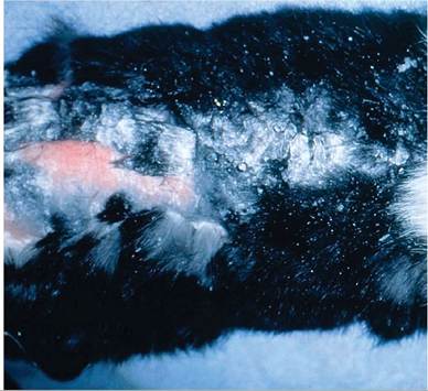
FIG. 6.82. Rabbit with chronic exfoliative dermatosis and sebaceous adenitis. Note the marked scaling lesions and alopecia over the dorsal aspect of the body (Source: M. Taylor and K.E. Linder.)
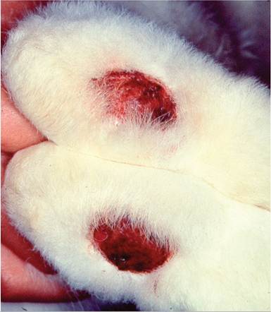
FIG. 6.83. Bilateral pododermatitis (sore hock) in an adult rabbit.
be associated with autoimmune hepatitis, thymoma, and cutaneous lymphoma in rabbits (see “Neoplasms”). Thus, it is important to pursue the possibility of the dermatosis having a paraneoplastic or neoplastic origin.
Pododermatitis: Sore Hocks
The plantar surface of rabbit feet is fully haired, but lesions consist of a circumscribed, ulcerated areas covered by granulation tissue and necrotic debris (Fig. 6.83). Purulent exudate may be adherent to the lesions. The problem is most commonly seen in heavy, mature adults. Poor sanitation, trauma from poor-quality, wire-bottom cages, and hereditary predisposition are factors that may influence the incidence of the disease. Staphylococcus aureus is the most frequent bacterium isolated from lesions.
Prolapse of the Deep Gland of
the Third Eyelid
Swelling and protrusion of the third eyelid has been associated with prolapse of the deep bilobed gland of the third eyelid. Affected animals present with a unilateral or bilateral protrusion of the third eyelid from the medial canthus of the eye.
Abnormal laxity of the connective tissue attaching the deep gland of the third eyelid to the bony orbital structures may be the underlying cause.Vertebral Fracture and Degenerative Spinal Disease
Posterior paralysis due to vertebral fracture or dislocation occurs all too often in domestic rabbits. The axial and appendicular skeletons of domestic rabbits are relatively fragile in proportion to their muscle mass. An unsupported, sudden movement of the hind limbs
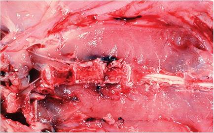
FIG. 6.84. Vertebral fracture in an adult rabbit due to improper restraint. Fractures typically occur in the lumbosacral region.
may exert sufficient leverage on the lumbosacral junction to cause a vertebral fracture. Depending on the duration of the problem prior to euthanasia and necropsy, the hindquarters may be soiled with urine and fecal material consistent with incontinence. The site of the fracture (or luxation) is usually the lumbosacral region (L7) (Fig. 6.84). There may be extensive hemorrhage in the underlying psoas muscles. Changes vary from luxation to multiple fractures of the affected vertebra, with extensive damage to the lumbosacral spinal cord. Degenerative changes of the nucleus pul- posis, usually involving the distal thoracic spinal segments, have been noted in rabbits as early as 3 months of age, with spondylosis among rabbits greater than 2 years of age.
Iatrogenic Nerve Damage
Following intramuscular injection of ketamine, xyla- zine, and acepromazine in the hind leg, rabbits were found to develop self-mutilation of digits. This syndrome was associated with inflammatory change and degeneration of the sciatic nerve in the region of the injections. Intrathecal injection of preservative-free ketamine has been shown to cause severe damage to the spinal cord and nerve roots, but ketamine with preservative did not. Long-term restraint of rabbits with hind leg extension was found to result in degeneration of sciatic nerves and skeletal muscle necrosis.
Affected rabbits had transient hind leg paresis.Tracheal Injury Following Intubation
Erosive to ulcerative tracheitis has been reported to occur in rabbits following tracheal intubation and inhalation anesthesia. The subjects were New Zealand White rabbits that were kept under general anesthesia for a period of 4-5 hours. Tracheal lesions have been identified in rabbits acquired from several different research facilities that had been subjected to this procedure. Clinically, stridor and mild cyanosis are the usual presenting signs in affected animals. At necropsy, changes may be confined to congestion of the tracheal mucosa. In more advanced cases, necrotic debris and blood are present
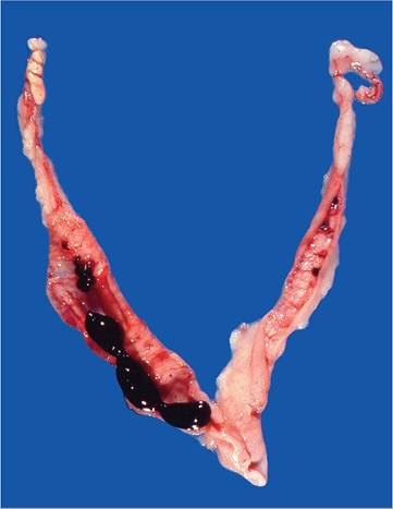
FIG. 6.85. Reproductive tract of a Californian breed doe with a
history of intermittent vulvar bleeding due to rupture of endometrial venous aneurysms. Blood clots are present within the uterine lumina.
on the tracheal mucosa. On histopathology, mucosal lesions are usually most extensive in the sections immediately distal to the larynx. Changes vary from multifocal mucosal ulceration to circumferential transmural ulceration of the tracheal mucosa.
Endometrial Venous Aneurysms
Multiple endometrial venous aneurysms have been associated with persistent urogenital bleeding. At necropsy, clotted blood is present in the uterine lumen (Fig. 6.85), and there are multiple blood-filled endometrial varices that consist of dilated, thin-walled veins (Fig. 6.86). These varices may rupture and bleed periodically into the uterine lumen, with subsequent hematuria. They have been observed in nonpregnant multiparous does. There is no evidence that predisposing factors, such as trauma or bleeding disorders, play a role in the disease.
Miscellaneous Genital Disorders
Cryptorchism may be occasionally encountered in bucks. In hot climates, bucks are prone to “seasonal infertility,” and seasonal involution occurs in the testes of some bucks during the nonbreeding season (see “Anatomic Features”).
Cystic endometrial hyperplasia is a relatively common disorder of aged does, and mucome- tra can be a sporadic finding. Because the uterine horns are completely separate with 2 cervices, mucometra may be unilateral or bilateral (Fig. 6.87).Urinary Sludge and Urolithiasis
As noted previously, intestinal absorption of calcium in rabbits occurs in direct proportion to dietary intake, as intestinal uptake is not influenced by vitamin D. Renal
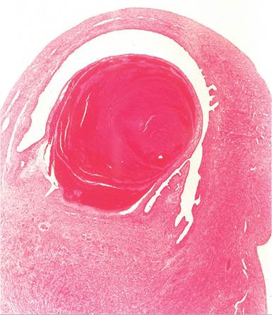
FIG. 6.86. Section of uterus from the previous figure. Note the marked dilation of the endometrial vessel and thrombosis.
calcium resorption and excretion are also in proportion to dietary intake, and regulated by parathyroid hormone, calcitonin, and vitamin D. When the resorptive capacity of the kidney is exceeded, calcium precipitates in the alkaline urine as calcium carbonate monohydrate, anhydrous calcium carbonate, and ammonium magnesium phosphate, resulting in the cloudy nature of rabbit urine in postweanling rabbits. When metabolic demand for calcium rises, the urine becomes less cloudy. When rabbits are fed high-calcium diets, such as diets rich in alfalfa, become dehydrated, or are suffering from other systemic disease, the likelihood of excess urinary mineral excretion takes on the characteristics of mud-like
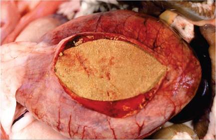
FIG. 6.88. Urinary bladder of a rabbit that died of urinary obstruction due to massive accumulation of intractable urinary sludge, which has the consistency of modeling clay. This material can accumulate rapidly.
“sludge.” This can happen very rapidly, resulting in acute urinary obstruction. The urinary bladder fills with intractable material that has the consistency of modeling clay (Fig. 6.88). Rabbits may also develop true uroliths in the renal pelves, ureters, urinary bladder (Fig 6.89), or urethra, resulting in hydronephrosis, hematuria, and obstructive uropathy.
Overweight and sedentary rabbits are at higher risk.Miscellaneous Kidney Disorders
Chronic renal disease, analogous to similar syndromes in aged rodents, is common in aged rabbits. Hydronephrosis may occur in association with urolithiasis. Fibrosis, with or without nephrocalcinosis, is common in rabbits over 10 months of age. Abscesses, pyelonephritis, and neoplasia (lymphosarcoma) may all involve the kidneys.
Amyloidosis
Amyloidosis occurs sporadically among rabbits, and is most likely to be observed in older rabbits that have been
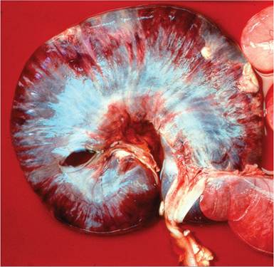
FIG. 6.87. Uterine horn of a doe with mucometra. The uterine wall has been incised, and the uterus is surrounded by serosanguinous fluid from the lumen. Because the uterine horns and cervices in the rabbit are anatomically separate, mucometra may be unilateral or bilateral.
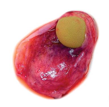
FIG. 6.89. Urolithiasis of the urinary bladder in a rabbit. (Source:
D. Imai, University of California, Davis, CA. Reproduced with permission from D. Imai.)
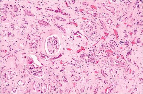
FIG. 6.90. Renal interstitial amyloidosis in a chronically immunized adult New Zealand White rabbit.
hyperimmunized or in rabbits with chronic infections, including pododermatitis and pyometra. Amyloidosis can also be readily induced experimentally. The kidney is the most commonly affected organ, in which amyloid deposits can be found in glomeruli, cortical interstitial tissue, or medulla (Fig 6.90). Mild early cases involve interstitial deposition in the medulla. Glomerular and cortical deposition tend to occur when medullary involvement is more severe. Medullary deposition may be associated with papillary necrosis or nephrolithiasis. Stomach, intestine, spleen, myocardium, adrenals, liver, lungs, and other organs may also be involved. Deposits in most organs are frequently perivascular, except spleen, in which deposits may be perifollicular or around the periphery of germinal follicles.
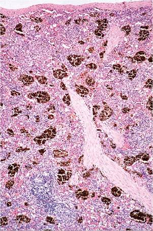
FIG. 6.91. Spleen of an adult rabbit with hemosiderin accumulation. This is a frequent incidental finding in aged rabbits.
Splenic Hemosiderosis
Aged rabbits tend to accumulate hemosiderin pigment in their spleens (Fig 6.91), which is probably due to excess iron in their diet.
BIBLIOGRAPHY FOR AGING AND MISCELLANEOUS DISORDERS
Tooth Abscesses
Tyrrell, K.L., Citron, D.M., Jenkins, J.R., Goldstein, E.J.C., & the Veterinary Study Group (2002) Periodontal bacteria in rabbit mandibular and maxillary abscesses. Journal of Clinical Microbiology 40:1044-1047.
Exfoliative Dermatosis and Sebaceous Adenitis
Florizoone, K. (2005) Thymoma-associated exfoliative dermatitis in a rabbit. Veterinary Dermatology 16:281-284.
Florizoone, K., van der Luer, R., & van den Ingh, T. (2007) Symmetrical alopecia, scaling and hepatitis in a rabbit. Journal of Veterinary Dermatology 18:161-164.
Prelaud, A.R., Jassies-van der Lee, A., Mueller, R.S., van Zeeland, Y.R.A., Bettenay, S., Majzoub, M., Zenker, I., & Hein, J. (2012) Presumptive paraneoplastic exfoliative dermatitis in four domestic rabbits. Veterinary Record 172:155.
White, S.D., Campbell, T., Logan, A., Meredith, A., Schultheiss, P., Van Winkle, T., Moore, P.F., Naydan, D.K., & Mallon, F. (2000) Lymphoma with cutaneous involvement in three domestic rabbits (Oryctolagus cuniculus). Veterinary Dermatology 11:61-67.
White, S.D., Linder, K.D., Schultheiss, P., Scott, K.V., Garnett, P., Taylor, M., Best, S.J., Walder, E.J., Rosenkrantz, W., & Yaeger, J.A. (2000) Sebaceous adenitis in four domestic rabbits (Orycto- lagus cuniculus). Veterinary Dermatology 11:53-60.
Prolapse of the Deep Gland of the Third Eyelid
Janssens, G., Simoens, P., Muylle, S., & Lauwers, H. (1999) Bilateral prolapse of the deep gland of the third eyelid in the rabbit: diagnosis and treatment. Laboratory Animal Science 49:105-109.
Vertebral Fracture and Degenerative Spinal Disease
Green, P.W., Fox, R.R., & Sokoloff, L. (1984) Spontaneous degenerative spinal disease in the laboratory rabbit. Journal of Orthopedic Research 2:161-168.
Jones, T., Lu, Y.S., Rehg, J., & Eckels, R. (1982) Diagnostic exercise: fracture of the lumbar vertebrae. Laboratory Animal Science 32:489-490.
Tracheal Injury Following Intratracheal Intubation
Nordin, U. & Lindholm, C.E. (1977) The vessels of the rabbit trachea and ischemia caused by cuff pressure. Archives of Otorhinolaryngology 215:11-24.
Nordin, U., Lindhom, C.E., & Wolgast, M. (1977) Blood flow in the rabbit tracheal mucosa under normal conditions and under the influence of tracheal intubation. Acta Anesthesiologica Scandi- navica 21:81-94.
Phaneuf, L.R., Barker, S., Groleau, M.A., & Turner, P.V. (2006) Tracheal injury after intratracheal intubation and anesthesia in rabbit. Journal of the American Association of Laboratory Animal Science 45:67-72.
Iatrogenic Nerve Damage
Mendlowski, B. (1975) Neuromuscular lesions in restrained rabbits. Veterinary Pathology 12:378-386.
Vachon, P. (1999) Self-mutilation in rabbits following intramuscular ketamine-xylazine-acepromazine injections. Canadian Veterinary Journal 40:581-582.
Vranken, J.H., Troost, D., de Haan, P., Pennings, F.A., van der Vegt, M.H., Dijkgraaf, M.G., & Hollman, M.W. (2006) Severe toxic damage to the rabbit spinal cord after intrathecal administration of preservative-free S(+)-ketamine. Anesthesiology 105:813-818.
Endometrial Venous Aneurysms
Bray, M.V., Weir, E.C., Brownstein, D.G., & Delano, M.L. (1992) Endometrial venous aneurysms in three New Zealand White rabbits. Laboratory Animal Science 42:360-362.
Urinary Sludge and Urolithiasis
Eckermann-Ross, C. (2008) Hormonal regulation of calcium metabolism in the rabbit. Veterinary Clinics of North America Exotic Animal Practice 11:139-152.
Amyloidosis
Hinton, M. & Lucke, V.M. (1982) Histological findings in amyloidosis or rabbits. Journal of Comparative Pathology 92:285-294.
Hinton, M. & Lucke, V.M. (1982) Ultrastructure of the kidney in amyloidosis of rabbits. Journal of Comparative Pathology 92:295-300.
Hofmann, J.R. Jr. & Hixson, C.J. (1986) Amyloid A protein deposits in a rabbit with pyometra. Journal of the American Veterinary Medical Association 189:1155-1156.
Miscellaneous Kidney Disorders
Hinton, M. (1981) Kidney disease in the rabbit: a histological survey. Laboratory Animals 15:263-265.
More on the topic AGING AND MISCELLANEOUS DISORDERS:
- BIBLIOGRAPHY FOR AGE-, MISCELLANEOUS-, ENVIRONMENTAL- AND DRUG-RELATED DISORDERS
- Radiculoneuropathy
- Chapter 14 ROUTINE TASKS AND DEALING WITH POISONS
- I OSTEOPOROSIS ^470 ^485 ^508 ^571
- CONGENITAL LUNG MALFORMATIONS
- Interstitial Lung Disease
- Chapter 50 Ovarian and Adnexal Disease