ANATOMIC FEATURES
The anatomy of the Syrian hamster has been reviewed by Bivin et al. (1987), Magalhaes (1968), and Murray (2012). Syrian hamsters have a characteristic compact body, short legs, and very short glabrous tails.
This is true for most other hamsters, with the exception of the Chinese hamster, which has a more elongated body and a longer tail. Hamsters possess 4 digits on the front feet and 5 on the rear feet. Syrian hamsters have remarkably abundant and loose skin. Adult female Syrian hamsters are larger than males. The female urethra has a separate opening from the vagina. Both sexes possess paired flank organs, which are most prominent in males. These organs consist of sebaceous glands, pigment cells, and terminal hair. They are darkly pigmented in mature males and appear to play a role in conversion of testosterone to dihydrotestosterone. The scent glands are located on the ventral midline in other species of hamsters. Hamsters have prominent depots of brown fat beneath and between the scapulae, in the axilla and neck, and around the adrenals and kidneys.Pathology of Laboratory Rodents and Rabbits, Fourth Edition. Stephen W. Barthold, Stephen M. Griffey, and Dean H. Percy. © 2016 John Wiley & Sons, Inc. Published 2016 by John Wiley & Sons, Inc.
The gastrointestinal tract has a number of significant features. As in mice and rats, the incisors (but not the molars) grow continuously (elodont). Many, although not all, genera of hamsters possess buccal pouches, which extend dorsolaterally from the oral cavity on either side of the shoulder region. These structures have been utilized as immunologically privileged sites, which allow xenograft transplants to survive. The utility of hamster cheek pouches as an experimental tool has been largely supplanted by immunodeficient mice. The esophagus enters the stomach at the junction of the nonglandular and glandular stomachs, which are divided by a muscular sphincter (Fig.
3.1). The non- glandular stomach, or forestomach, features an elevated pH and a complex microbiome, which contribute to fermentative digestion. Paneth cells are a normal constituent of small intestinal crypts. The cecum is divided into apical and basal portions separated by a semilunar valve. In fact, there is a series of 4 valves in the ileoce- cocolic region of the hamster. As with other rodents and rabbits, hamsters rely on coprophagy for nutrition. The liver is divided into 4 lobes, with a gall bladder. As in the mouse and rat, intranuclear cytoplasmic invagination (inclusions) and eosinophilic cytoplasmic inclusions in hepatocytes can be found, particularly in diseased livers.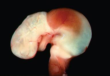
FIG. 3.1. Stomach from a hamster, depicting the nonglandular and glandular regions. Note the esophageal entry at the junction of each.
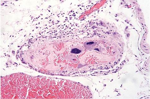
FIG. 3.2. Trophoblast cells within mesometrial vessel in a pregnant hamster.
The respiratory tract is similar to that of other laboratory rodents, with no respiratory bronchioles. Lungs have a single left lobe and 5 lobes on the right side (cranial, middle, caudal, intermediate, and accessory). As desert animals, Syrian hamsters have water-conserving kidneys with elongated single papillae that extend into the ureters. The female reproductive tract consists of a duplex uterus with 2 cervical canals that merge into a single external cervical os. There are 7 pairs of mammary glands. The male testes and accessory glands, as with most rodents, are comparatively large and prominent. Adult males develop large adrenal glands, due to enlargement of the zona reticularis to 3 times the size of females. This enlargement is related to season and sexual maturity. The hamster placenta differs somewhat from the hemochorial placentation of other laboratory rodents and is termed “labyrinthine hemochorial.” Trophoblastic giant cells of the fetal placenta are in direct contact with the maternal bloodstream and tend to migrate within the maternal vasculature. They have a tropism for arterial blood and can be found inside uterine vessels in the mesometrium (Fig. 3.2). They can persist for up to 3 weeks postpartum. On occasion, trophoblasts may be found in the pulmonary vessels of pregnant females.
Erythrocytic polychromasia is relatively common in hamsters, with moderate anisocytosis. Erythrocyte life spans vary from 50 to 78 days. Life spans are increased during hibernation. Leukocyte counts are 5,000-10,000/ml. Approximately 60-75% of the circulating leukocytes are lymphocytes. Neutrophils have densely staining eosinophilic granules and thus may be referred to as heterophils.
More on the topic ANATOMIC FEATURES:
- ANATOMIC FEATURES
- References
- Bibliography
- Skeletal System
- Constrictive Pericarditis
- Screening for Fetal Abnormality in Multiple Pregnancy
- Postoperative Complications and Postoperative Emergencies
- CHAPTER 38 ASPERGILLOSIS
- 18 Autoimmune Disease in Pregnancy
- 40 Abnormal Uterine Bleeding