26 Inflammatory bowel disease and adverse reaction to food in a dog
Initial presentation
Six months of soft to watery diarrhoea and weight loss and episodes of vomiting
Signalment: 4-year-old entire male Sharpei, body weight 15.5 kg
Case history
The dog presented with a chronic, 6-month history of diarrhoea and weight loss.
He was producing large volumes of soft faeces, with no increase in frequency of defecation, no blood or mucus in the faeces and no tenesmus while defecating. There had been episodes of vomiting occurring nearly every day. The dog usually vomited digested food and bile and this often occurred soon after eating. The owner reported that the dog had become quieter than he had been previously.Clinical tip
The presence of weight loss, large volumes of faeces and lack of tenesmus, mucus or haematochezia indicate diarrhoea of small intestinal origin. Localizing the source of the diarrhoea aids the clinician in making a differential diagnoses list and deciding upon diagnostic techniques.
The presence of digested food and bile in the vomited material indicates that it is likely to be true vomiting rather than regurgitation. The length of time after eating that an event takes place does not necessarily indicate whether it is vomiting or regurgitation.
The dog had been fed a variety of diets in an attempt to control the gastrointestinal signs, including a highly digestible diet for gastrointestinal disorders and a premium dry dog food.
The current diet being fed was a commercial dry diet based on lamb and rice. The dog had a good appetite while on this food, but there was no improvement in the diarrhoea or vomiting. In addition to eating dog food, the dog also scavenged from rubbish and was fed treats of human food.
His vaccinations were upto date and he had been treated for parasites 6 months prior to presentation. He had not travelled outside of the UK.
The diagnostic tests and treatments from the previous veterinary surgeon included:
• Faecal examination for parasites: negative
• Treatment with sulfasalazine and metronidazole for 1 week (doses not reported)
• Dietary therapy: a dry veterinary diet designed for intestinal disease for 2 weeks, then a dry commercial premium dog food for 2 weeks
There had been an initial improvement on these treatments and then the diarrhoea reoccurred 2 months later.
He was treated again with sulfasalazine, metronidazole (doses not reported) and prednisolone at 0.6 mg/kg, with no improvement.Sulfasalazine is used for large intestinal diarrhoea so would not be expected to improve the signs in this dog. Metronidazole does help in some cases of diarrhoea. Prednisolone should probably not be used without a confirmed diagnosis; if it was being used for presumptive inflammatory bowel disease, the dose was too low as it is between the physiological dose for dogs (0.15-0.25 mg/kg/day) and the anti-inflammatory dose for dogs (0.5-1 mg/ kg/day).
Clinical tip
While the diets that were used in this case are highly digestible they do include several protein sources and are not appropriate as elimination diets. The first ingredient in the lamb and rice diet was wheat, showing that it is necessary to read the labels. The diets at the time of this case contained the following main ingredients:
a Veterinary intestinal diet: maize, gluten meal, chicken and turkey meal
b Premium dry diet: chicken, rice, maize, poultry meal, gluten meal, wheat, fish oil, egg
c Lamb and rice based diet: wheat, lamb, rice, bran
On physical examination the dog was quiet and alert and his hydration was adequate. His body condition score was 3/9, with evidence of muscle loss. His mucous membranes were pink and capillary refill time was fungal
• gastrointestinal parasites
• inflammatory bowel disease
• infiltrative neoplasia, e.g. lymphoma, mastocytosis
• partial obstruction, e.g. intussusception, foreign body
• brush border defects
• lymphangiectasia
• Pancreatic disease
• chronic pancreatitis
• exocrine pancreatic insufficiency
• pancreatic neoplasia
• Liver disease
• bile duct obstruction
• liver failure
• Kidney disease - unlikely as he did not have polyuria/polydipsia (pu/pd), although glomerular disorders may present without pu/pd
• Miscellaneous
• congestive heart failure - no signs consistent with this
• immunodeficiencies - e.g.
IgA deficiency, which has been reported in the Sharpei dog• hypoadrenocorticism
Weight loss with good appetite
• Dietary
• unbalanced or poor quality diet
• insufficient food provided
• Malassimilation
• intestinal disorders as above for diarrhoea
• exocrine pancreatic insufficiency
• liver failure
• Nutrient loss
• diabetes mellitus (unlikely as not pu/pd)
• protein losing enteropathy
• protein losing nephropathy
• Increased nutrient requirements
• fever (not present)
• hyperthyroidism (unlikely)
Occasional vomiting
• Dietary, small intestinal, liver and pancreatic disorders as above
• Stomach
• gastritis
• gastric ulcers
• inflammatory bowel disease
• foreign body
• parasites
• Systemic disease
• uraemia
• liver failure
• sepsis
• congestive heart failure
• acidosis
• hypoadrenocorticism
• ketoacidotic diabetes mellitus
• hyperparathyroidism
• gastrinomas
• Neurological diseases (no other signs consistent with these in this dog)
• dysautonomia
• vestibular disease
• CNS disease
• Drugs, toxins (no known history outside of medications provided by referring veterinary surgeon)
Case work-up
Minimum data base
Haematology, serum chemistry and urinalysis were performed. The most significant findings were a hypoalbuminaemia of 15.2 g/l (reference range 26-35 g/l) and borderline hypoglobulinaemia of 21.9 g/l (reference range 22-45 g/l). He also had a peripheral eosinophilia of 1.7 ? 109∕l (reference range 0-1 ? 109/l). His urine had a specific gravity of 1.040 with a pH of 6.5. The urine protein to creatinine ratio was 0.8 (slightly elevated above the reference range of 0.5) and there were calcium oxalate crystals present in the sediment.
Low albumin may be caused by intestinal loss, urinary loss, decreased liver production due to liver disease, third spacing, a shift from the vascular space into the interstitial space or decreased liver production during an acute phase reaction from inflammation.
With a concurrent decrease in globulins and only a mild increase in urine protein and a history of diarrhoea, intestinal loss was the most likely diagnosis in this dog.Eosinophilia may be caused by hypersensitivity (e.g. food, parasites), eosinophilic diseases (eosinophilic inflammatory bowel disease, eosinophilic pneumonitis, eosinophilic granulomatous disease, eosinophilic leukaemia) and is also sometimes seen with mast cell tumours, carcinoma and hypoadrenocorticism.
Faeces were submitted for parasitology and culture of enteropathogens; both of these results were negative.
Clinical tip
Many intestinal parasites shed eggs only intermittently, so a negative faecal examination does not rule out parasites. Doing three faecal examinations from three different stool samples has been recommended especially in cases with gastrointestinal signs, de-worming is usually recommended.
Adrenocorticotrophin hormone stimulation test
An adrenocorticotrophin hormone (ACTH) stimulation test was performed and the results were within the reference range (pre-ACTH stimulation of 223 mmol/l increasing to a post-stimulation of 443 mmol/l), ruling out hypoadrenocorticism.
Endoscopy
Endoscopy showed an increased duodenal granularity, hyperaemia and friability (Fig 26.1). Results of the histopathology of the gastric and small intestinal biopsies showed an increased in filtration of eosinophils, consistent with eosinophilic inflammatory bowel disease.
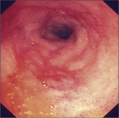
Fig 26.1
Endoscopy picture of the small intestine of a dog with eosinophilic inflammatory bowel disease. There is roughening of the mucosa with small pinpoint haemorrhagic areas
Clinical tip on inflammatory bowel disease scoring
A scoring index for canine inflammatory bowel disease (IBD) (the canine IBD activity index or CIBDAI) has been developed. The canine IBD activity index includes:
A. attitude/activity
B.
appetiteC. vomiting
D. stool consistency
E. stool frequency
F. weight loss
These six variables are scored 0 to 3:
0 = normal
1 = mild change
2 = moderate change
3 = severe change
The summary of these six variables gives the total composite CIBDAI score, which is interpreted as:
0 to 3 = clinically insignificant disease
4 to 5 = mild IBD
6 to 8 = moderate IBD
9 or greater = severe IBD
The CIBDAI has shown correlation with the acute phase protein reactant C reactive protein and severity of histology changes. Following treatment for IBD both the CIBDAI and serum C reactive protein decreased.
To calculate the CIBDAI score in this dog the following were used: a mild decrease in his activity (1), no change in his appetite (0), a moderate change in his faecal quality (2) moderate amount of vomiting (2) and moderate weight loss (2), for a score of 7 or moderate IBD.
Treatment and outcome
This dog was initially treated with prednisolone at 2 mg/kg po q 24 hours, subcutaneous cobalamin injections of 500 μg per week for 4 weeks and fenbendazole at 50 mg/kg po q 24 hours for 3 days. The owner did not wish to change the food, so he continued to feed the commercial lamb and rice diet.
At 19 days, there was no response to treatment and the dog had vomited after being fed pancakes. Prednisolone was continued and the dog was changed on to a single novel protein, gluten-free, highly digestible diet.
At 37 days, the dog was still occasionally vomiting and the stools were loose. The owner said he had not been giving the prednisolone consistently.
At the day 42 check up, the owner had been giving prednisolone, but had added potatoes, gravy, bread and poultry to the diet and there was no improvement in the clinical signs. The prednisolone dose was decreased to 1.6 mg/kg po q 24 hours.
At the day 56 check up, the owner reported that he had complied with the dietary and medication therapies. The dog had gained 1 kg and his stools were soft but formed.
The prednisolone dose was decreased to 1.25 mg/kg po q 24 hours and a therapeutic plan was made to decrease the dose about 20% every 3 weeks, until the dose was less than 0.5 mg/ kg q 24 hours, when it was to be changed to an every other day regime.At the day 120 check up, the owner reported that he had stopped giving prednisolone. The dog had gained 2.5 kg and the stools were still soft to firm and he was much brighter and more active. The serum albumin and globulins were within the reference range.
Discussion
This dog did not improve until wheat products were eliminated from the diet and the medical treatment (prednisolone) did not work without dietary intervention.
Because of the response to an elimination diet, it is highly likely that this dog had an adverse reaction to food. In order to determine what ingredient caused the reaction and to confirm that the response was due to an adverse reaction to food, a food challenge would have been necessary.
Clinical tip
Elimination-challenge dietary trials confirm or rule out adverse reactions to food, but do not establish an immune mediated basis (hypersensitivity) for the reaction. It is however, probably not important to establish an immune mediated basis as it does not change the management of the case.
The elimination trial involves putting the patient on a restricted diet of a single protein source and a single carbohydrate source. The diet used should contain ingredients to which the animal has not previously been exposed. There is no universal ‘hypoallergenic diet’ containing intact proteins, as allergenicity depends upon the foods that an individual has previously been fed. Homemade diets are often recommended; however, 20% of the dogs having no signs when fed a homemade diet show signs when fed a canned diet with the same constituents. Another option is to use a commercial diet with hydrolyzed protein. Supplements with n-3 or n-6 fatty acids should not be given, as they may cause allergic reactions in animals that are allergic to fish.
In a study on food sensitive cats with gastrointestinal signs there was an immediate cessation of vomiting and a decrease in diarrhoea within 3 to 4 days, in most of the cats after feeding a novel protein diet. Most of these cats also showed a recrudescence of signs after 3 to 5 days of an antigenic challenge.
After a 2-week period with no clinical signs, a small amount of a second protein source (e.g. one-half to two tablespoons of powdered milk) to which the animal has been previously exposed can be added to the controlled diet and fed for 3 days - or less if signs are observed. This procedure is repeated weekly until a suspect food appears to cause the signs. After the suspect food is identified, remove that protein source from the diet and observe for evidence of resolution of signs. After 1 to 2 weeks of no signs, challenge the animal with the suspect food again.
This owner, as are many, was reluctant to challenge the animal after the resolution of signs and it was necessary to design a diet without confirmation of the allergen. The diet fed in cases like this should be palatable, complete and balanced. Use of a high quality commercial gluten free, moderate to low fat, lactose free diet with limited antigens and few additives may work well in these cases. In some cases it is difficult to find a protein source which has not been fed; in these cases use of a hydrolyzed protein diet or one which has not been fed for the previous 6 months may be tried. While many of these patients are underweight, as was this one, excess dietary fat should be avoided during small intestinal malassimilation as malabsorbed fatty acids and bile acids can cause secretory diarrhoea.
It may have been possible to manage this case with dietary therapy alone; however, most cases of eosinophilic inflammatory bowel disease also require therapy with immune suppressive drugs such as prednisolone.
The lack of owner compliance with diet and medications in this case complicated the management and prolonged the time to recovery. Many medications are not given according to directions and clinicians must be sensitive to the owners’ ability and willingness to administer medications and control the diet. Asking clients in a non-judgmental manner if they are having any problems giving the prescribed medication or if they have missed any doses is one way to assess compliance and identify any difficulties clients may have in giving medications to pets. If a client does not admit to missing any doses but compliance is still in doubt, a follow-up appointment for re-examination and a pill count may reveal a problem with compliance.
Prognosis
In one retrospective study, only 26% of 80 cases of IBD were considered to be in remission at a 6-month follow-up. Many of the dogs (about 50%) showed intermittent signs of disease and most of these (65%) were still on treatment. The presence of hypoalbuminaemia is a negative prognostic indicator and an elevated canine pancreatic lipase concentration is also associated with a negative outcome.
More on the topic 26 Inflammatory bowel disease and adverse reaction to food in a dog:
- Inflammatory Bowel Disease
- Canine inflammatory bowel disease activity index (CIBDAI)
- 39 Colonic inflammatory bowel disease in a cat
- 15 Lymphocytic inflammatory bowel disease/al- imentary lymphoma in a cat
- Adverse Food Reactions (AFR)
- Pelvic Inflammatory Disease
- Costs of group living include greater energy expenditures, more competition for food, and higher risks of disease
- Congenital malformations of lower gastrointestinal tract may be divided into three categories—(a) small bowel malformations, e.g. atresia, malrotation, etc., (b) colorectal malformations, e.g. Hirschsprung disease or anorectal anomalies, and (c) abdominal wall defects with gut herniation.
- Bowel Management
- ADVERSE EVENTS FOLLOWING IMMUNIZATION (AEFI)