LOUPING-ILL
HUGH W. REID
The Moredun Foundation, Pentlands Science Park, Bush Loan, Penicuik, Midlothian, Scotland
Louping- ill is a viral disease that has been recognized in domestic animals and, rarely, humans for many years but has also been established as a significant cause of mortality in wild red grouse (Lagopus lagopus scoticus).
The cause is a flavivirus belonging to the closely related TBE complex of viruses transmitted by ixodid ticks and distributed throughout the northern temperate regions.
Initially louping-ill was described as a condition of domestic sheep in the UK, but a similar disease of sheep has been found in Norway, Spain and Bulgaria, while cattle, horses, pigs, South American camelids, dogs and humans have been affected in the UK.
Infection of humans is only rarely diagnosed, but disease is relatively common in all categories of domestic livestock as well as wild red grouse exposed to the virus.
The pathogenicity of Louping-ill virus for red grouse resembles that of a virus exotic to the habitat of the red grouse. However, red grouse are one of the few vertebrate species almost unique to the UK, as is the louping-ill virus. Before the early 1400s, the upland rough pastures in northern Britain were largely used as summer pastures for cattle. The replacement of this traditional practice with large-scale sheep-raising probably enhanced the host potential for I. ricinus ticks, effectively introducing the species to this habitat. Furthermore, abundance of alternative hosts would have been meagre, and ticks would have predominantly relied on sheep for blood meals. It is speculated that these changes facilitated the adaptation of an ancestral TBE virus, which had either been previously maintained in a forest/woodland ecosystem in Britain or was introduced de novo from continental Europe. Over the next 400 years Louping-ill virus evolved to this specific habitat and was progressively dispersed across the British Isles.
The red grouse is a highly specialist feeder with a predilection for heather ( Calluna vulgaris). Molecular analysis of the envelope gene of approximately 50 isolates confirms that Louping-ill virus has evolved from an ancestral TBE virus over approximately 400 years. There is thus excellent agreement between the historical appraisal of the probable evolution of Louping-ill virus and molecular analysis of contemporary isolates.
As transmission of Louping-ill virus in nature is dependent on the vector tick Ixodes ricinus, factors determining the activity and distribution of the tick dictate the epidemiology of louping-ill. Virus is transmitted trans-stadially and there is no evidence of transovarial transmission. Ticks become infected when they ingest viraemic blood, but relatively high titres of virus are required to establish infection. Thus only nymphs and adults that have derived their blood from a susceptible viraemic host as larvae and nymphs, respectively, are capable of transmitting virus.
To determine which vertebrates might contribute to amplifying the virus, a series of experimental infections in candidate domestic and wild species was undertaken to establish the titres of virus that developed in blood. The only mammalian species that consistently developed virae- mias of a sufficient intensity to infect ticks were domestic sheep, in which viraemias exceeded threshold levels to infect ticks for 1 to 3 days. Similar experiments established that red grouse also developed sustained viraemias substantially above threshold levels to infect the tick. However, approximately 80% of infected birds died with a florid virus-induced encephalitis.
Subsequent experiments also established that mountain hares (Lepus timidus) could provide infected blood meals through a mechanism called co-feeding, i.e. ticks feeding on adjacent areas of skin to an infected tick could acquire infection through local transmission of virus in interstitial fluid in the absence of viraemia.
These studies suggested that the only essential maintenance vertebrate host of louping-ill viruses was domestic sheep, whereas infection of other species was tangential and only makes a minor contribution to virus maintenance. Field studies have supported this view and, where louping-ill virus becomes established, it is responsible for heavy mortality among red grouse, with populations declining and often disappearing1-20).
Field studies of grouse suggested that the numbers of ticks parasitizing grouse was relatively low and could not explain the high incidence of louping- ill in red grouse. Examination of grouse droppings indicated that grouse consumed numerous ticks, and experiments confirmed that grouse could readily be infected by the oral route. It was concluded that the majority of grouse became infected through ingestion of infected ticks, a route of transmission that accounted for up to 95% of infections1-21).
Following inoculation of virus, replication occurs in the lympho-reticular system over 4 to 5 days. This is accom-
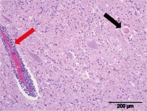
FIGURE 9.5 Histological section of the brain of a sheep with clinical louping-ill. Note the characteristic perivascular cuff comprised of lymphoid inflammatory cells (red arrow) and necrotic neurons (black arrow). Haematoxylin and eosin stain. Courtesy of Dr Mark Dagleish (Moredun Research Institute).
panied by minimal clinical manifestations, although a febrile response and some malaise may occur. It is during this phase of the disease that animals become viraemic and virus may invade the CNS. Decline and elimination of viraemia is associated with the appearance of neutralizing and haemagglutination-inhibiting antibody, which is maintained for life in survivors. Virus persists in the brain for a few days following elimination of infectivity from the periphery and lesions characterized by a non-suppurative encephalomyelitis and neuronophagia develop1-22) (Figure 9.5).
Depending on the species involved, a variable proportion recover.Louping-ill is an RNA virus that can be isolated readily in tissue culture. However, virus is most usually detected today by specific RT-PCR(23). IHC labelling of nonsuppurative encephalitic lesions if combined with PCR detection of viral nucleic acid are diagnostic. Of great value in epidemiological investigation of this disease in wildlife is serology, and this has been used on red grouse and brown hare among other wild species in the UK to monitor the efficacy of the strategies mentioned below. Though there have been several attempts to apply ELISA tests for the detection of antibody, the HI test(24) has been found to be consistently reliable across a spectrum of domestic and free-living mammals and birds.
Sustained control strategies designed to suppress ticks and virus circulation through intensive acaracide treatment of sheep, vaccination of all sheep and reduction of alternate wild animal tick hosts, where required, have resulted in a substantial reduction in the prevalence of louping-ill virus and the recovery of grouse populations(25).
ACKNOWLEDGEMENTS
The author acknowledges the contribution to the manuscript and the figures kindly provided by Dr Mark Dagleish (Moredun Research Institute).
More on the topic LOUPING-ILL:
- LOUPING-ILL
- 3 SELECTED SOCIO-ECONOMICALLY IMPORTANT WILDLIFE RELATED PATHOGENS AND DISEASES IN EUROPE
- 2 SELECTED ZOONOTIC PATHOGENS WITH EUROPEAN WILDLIFE RESERVOIRS/HOSTS
- 5 Appendices