Otitis Externa
Otitis externa is a very common problem in the dog. It is seen less often in the cat. The prevalence of disease in the dog has been reported to be between 10% and 20% of total patient admissions, with the prevalence in the cat being 2% to 10%.
Many factors may predispose an animal to ear disease, including allergy, disorders of kera- tinization, parasites, foreign bodies, autoimmune diseases, treatment errors, nutritional factors, hormonal factors, and any process that interferes with epithelial migration and desquamation, including benign and malignant tumors. All of these factors serve to alter the microenvironment of the ear canal.Successful treatment relies on proper diagnosis and correction of any primary, predisposing, or perpetuating factors. It is important to evaluate and correct, if possible, any alterations in the microclimate of the external ear canal. The normalization of the microclimate aids in keeping an ear disease free, after all other factors have been identified and addressed.
Structure of the External Ear Canal
The epidermis of the ear canal is similar in structure to that of the interfollicular epidermis of the skin. However, from a gross anatomic standpoint, this epidermis is rolled into a tube, forcing the glandular secretions of the sebaceous and apocrine glands into a canal instead of to the skin, where the secretions can be more easily removed by the normal keratinization and desquamation process of the epidermis. Further modifications of the anatomy such as pendulous ears or excessive ear canal hair result in significantly more otitis externa than that seen with other ear types. Dogs with erect ears, regardless of the amount of ear canal hair, have less risk of developing otitis externa than mongrel dogs. In humans and in the guinea pig, and presumably in the dog and cat, the superficial epidermis and keratinized stratum corneum migrate laterally from the tympanum.
This process keeps the proximal ear canal and tympanum free from cerumen and debris.Microclimate of the External Ear Canal
The principal factor affecting the microflora within the external ear canal is the microenvironment. In several studies the temperature within the external ear canals of dogs measured between 38.2° to 38.4° C (100.7° to 101.1°F). There was no significant difference between breeds of dog or whether the pinnae were pendulous. The temperature within the external ear canal rises if otitis externa is present, to a mean of 38.9° C (102° F). Even when environmental temperature increased, there was only a small rise in the temperature of the external ear canal, illustrating how the environment within the ear canal is buffered to a degree from the external environment.
The relative humidity of the external ear canal in one study of 19 dogs was 80.4%. This was stable through the day, rising only 2.3%, compared with the 24% rise in the humidity of the external environment. However, this increase in average humidity from a relatively high baseline may predispose the canal epithelium to becoming overhydrated and macerated, creating a more ideal environment for bacterial proliferation.
The pH of the external ear canal in normal dogs is 4.6 to 7.2, with a mean of 6.1 in males and 6.2 in females. The pH is seen to change in otitis externa, with a mean of 5.9 (range 5.9 to 7.2) in acute cases and 6.8 (range 6.0 to 7.4) in chronic cases. The data from this study were analyzed by another author, who showed that in cases of otitis externa associated with Pseudomonas spp. the pH was significantly higher (mean of 6.85) than in cases of otitis externa in which no Pseudomonas was isolated (mean of 5.7). Thus pH either seems to play an important role in the predominant type of bacteria colonizing the ear canal, or the bacteria and its products alter the pH of the environment.
Cerumen coats the lining of the external ear canal. It is composed of lipid secretions from the sebaceous gland and apocrine (ceruminous) glands mixed with sloughed epithelial cells.
The lipid content of cerumen from a normal ear canal of a dog can vary widely (18.2% to 92.6%), as does the type of lipid within the cerumen. The types of cerumen lipids found in normal dog ears were cholesterol, 100%; cholesterol esters, 93.8%; free fatty acids, 93.8%; fatty aldehydes, 93.8%; waxes, 93.8%; triglycerides, 68.8%; lecithin, 56.3%; and sphingomyelin, 18.8%. The methodology used in the study was not able to detect small amounts of lipids; therefore this list of lipids accounts for only the major lipids found in the canine ear canal.1The total amount of lipid in the cerumen also varies considerably in dogs with otitis externa (4.3% to 69.6% of cerumen) and is significantly lower than that from healthy ears. This may be due to pathologic changes in the glands responsible for the formation of cerumen. The apocrine glands are thought to maintain the consistency of the cerumen, although they probably contribute little in the way of lipids. Lipids excreted by sebaceous glands probably constitute a large proportion of cerumen lipids. In chronic otitis externa the apocrine glands become hyperplastic and cystic, whereas the sebaceous gland can vary from hypertrophic to atrophic. This may cause pathologic changes of the cerumen in otitic ears, producing a generally lower lipid yield. The high lipid content of normal cerumen helps maintain normal keratiniza- tion of the epidermis and aids in the capture and excretion of debris that is produced within. The high lipid content also results in a relatively lower humidity within the lumen of the ear canal. With a decrease in secretion by the sebaceous glands or a dilutional effect by increased production by the apocrine glands, humidity within the ear canal rises, and maceration, followed by otic inflammation and infection, results.
Microbiology of the External Ear Canal
Common diagnostic tests used to evaluate an animal with ear disease include otoscopy, cytology, and bacterial culture and sensitivity.
Since the normal ear canal is not a sterile environment, cytologic results, as well as results from culture and sensitivity testing, must be viewed in conjunction with information obtained from a thorough otoscopic examination. On otoscopic examination of the normal ear canal, a small amount of yellowish-brown wax may be seen. There should be no erosion, ulceration, or inflammation of the epidermal lining. The ear canal should be able to accommodate an average-size otoscopic cone without undue pressure on the sides of the canal. The tympanic membrane should be easily visualized in a willing patient.A cytologic examination of exudate from the ear of a patient with otitis externa should be performed during each examination. Samples can be taken with a dry, cotton-tipped swab placed as far into the canal as is comfortable and safe—usually to the junction of the vertical and horizontal canals. Obtained exudate is placed on a slide and mixed with mineral oil to examine for ear mites. A second sample should be rolled on a slide and stained with either a Wright-Giemsa stain, modified Wright- Giemsa stain, or Gram’s stain. If the exudate is greasy or waxy, heat fixation before staining may be helpful. The stained slides should be examined with the low- (100?) and high- (1000?) power objectives of the microscope. A low-power scan permits rapid evaluation of the smear and identification of the best area(s) of the slide for high-power examination. Otic parasites will be seen at low power (Figure 9-1). High-power oil immersion (1000?) evaluation permits quantitation of yeast and examination for bacteria.
Smears should be evaluated for (1) the number and morphology of bacteria, (2) the number of yeasts, (3) the presence of fungal elements, (4) the presence of parasites, (5) the number and types of leukocytes and whether they contain phagocytized microorganisms, (6) the presence of excessive cerumen, (7) the presence of excessive keratinaceous debris, and (8) the presence of neoplastic cells.
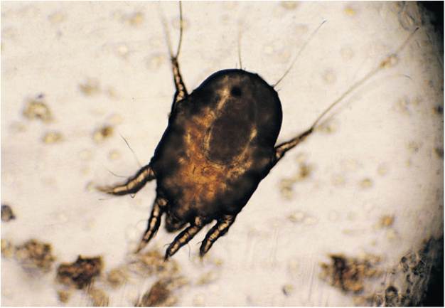
Figure 9-1
Gravid female Otodectes cynotis from a dog ear smear (magnification 100?).
The physical characteristics of the material collected from the external ear canal may provide clues as to the underlying cause. The normal ear canal should have small quantities of yellowish to light brown nonodiferous ceruminous discharge. Dark yellow to light brown discharge is seen in ears infected with gram-positive cocci. Pale yellow, thick, sweet, or sewer-smelling purulent exudates are noted in ears infected with gram-negative rods. Copious, dark brown, waxy, or “yeasty” smelling exudates are seen with the yeast Malassezia canis. Dark brown or black, crumbly exudate resembling coffee grounds suggests the presence of the parasitic mite Otodectes cynotis. A white exudate with no odor, resembling melted candle wax, is often seen after resolution of infection in cases of chronic otitis externa. There is no evidence cytologically of any microorganisms in this exudate, just continued excessive oily or waxy cerumen of hyperplastic ears. Combinations of the various etiologic agents lead to alterations of the characteristics listed previously.
On microscopic evaluation, gram-positive cocci, most often staphylococci, occur singly, in pairs, or in short chains. Streptococci will also be gram positive, but the cocci are usually smaller. Streptococci do not commonly form chains when observed in smears from ear infections. Medium sized gram-negative rods are most likely to be Pseudomonas, Proteus, or Escherichia coli. Small beaded or club-shaped grampositive rods are likely to be Corynebacterium. The presence of large gram-positive rods in gram-stained smears suggest the presence of Bacillus or its anaerobic counterpart Clostridium. Malassezia canis occurs as an oval-shaped yeast in which the buds are broad based. As a result of budding the M. canis often appear peanut-shaped.
In the Gram’s stain, M. canis is gram variable.The normal cytology of the external ear canal is characterized by the presence of squamous epithelial cells and low numbers of commensal but potentially pathogenic microorganisms, including M. canis and Staphylococcus intermedius. There is some debate as to whether M. canis is a primary pathogen; however, it is found three times more frequently in ears with otitis externa than in normal ears. M. canis is the most common organism demonstrated in ear specimens. It has been isolated in up to 49% of normal dogs and 23% of normal cats. In dogs, M. canis has been isolated in up to 80% of otitis externa cases and is probably the most common complicating factor of allergic otitis. In one study it was proven that M. canis in the presence of extraneous influences of atraumatic manipulation or moisture enhanced the conditions necessary for proliferation of the organisms and gross and microscopic evidence of otitis externa, establishing this organism as an opportunistic pathogen.2 On cytologic examination, there are usually few to no leukocytes unless bacteria are also present with the broad-based budding yeasts.
The significance of numbers of M. canis or bacterial organisms found on cytologic evaluation varies among authors. Few pure quantitative studies concerning populations of bacteria and yeast residing in the external ear canal are published in the veterinary literature.
A semiquantitative cytologic evaluation of the exudate from normal ears and from ears with otitis externa in the dog and cat has been performed.3 In this study it was shown that numbers of cornified squamous cells are not consistently correlated with clinical findings, as animals with high counts may not show any clinical sign of otitis externa. In the above mentioned study, the results indicated that two or fewer M. canis yeast cells per high-power dry field (400?) should be considered normal in the dog or cat. Mean counts of greater than or equal to five yeast cells per high- power dry field in the dog and greater than or equal to 12 yeast cells per high-power dry field in the cat should be considered abnormally increased. It was theorized that Malassezia otitis may be a more common clinical entity in the cat than previously reported in the literature and that clinical signs are associated with higher mean yeast counts than in the dog.3
In reference to bacteria, mean counts per high-power dry field (400?) less than or equal to five bacteria per field in the dog and less than or equal to four bacteria per field in the cat should be considered normal, whereas mean counts greater than or equal to 25 bacteria per field in the dog and greater than or equal to 15 bacteria per field in the cat were abnormally increased.3 Degenerating neutrophils are seen most often with significant bacterial disease and may indicate the need for systemic antibiotic therapy.
Relying on bacterial culture alone without considering the results of cytologic evaluation to determine the significance of bacteria or yeast in an inflamed ear may lead to inappropriate use of antimicrobials, because normal and acutely infected ears may harbor the same organisms. It is the opinion of many authors that cytologic evaluation of otic exudate provides greater diagnostic information about the significance of bacteria and yeast in ear disease than do culture results, especially since it is unlikely that pathogenic organisms will be cultured if they are not seen on cytologic examination. However, cultures should be considered in recurrent or refractory cases, especially those involving gram-negative bacteria and when otitis media is suspected.
The most common gram-positive organism isolated from cases of canine otitis externa is Staphylococcus intermedius (Figure 9-2). Other gram-positive bacteria isolated include Streptococcus spp., Micrococcus spp., Staphylococcus aureus, Staphylococcus epidermidis, Corynebacterium spp., and Bacillus spp. Gram-negative organisms isolated from cases of canine otitis externa include Pseudomonas spp., Proteus spp., Klebsiella spp., E. coli, and Pasteurella spp. These gram-negative organisms, particularly Pseudomonas, are seen more commonly in chronic cases of otitis externa (Figure 9-3).
Pseudomonas aeruginosa is perhaps the most difficult to manage of the bacteria that infect the ear canal. Pseudomonas is intrinsically insensitive to many antimicrobial drugs because of the low rate of passage of antibiotics across its outer membrane. There is evidence accumulating that P. aeruginosa isolates from dogs with otitis externa are becoming resistant to a number of antibacterial agents, including the fluoroquinolones. Pseudomonas thrives in an environment created by chronic inflammatory changes of the ear canal.
The most common yeast agent cultured from canine ears is M. canis (Figure 9-4). Other yeast organisms isolated from cases of canine otitis externa include Candida, Cryptococcus, Rhodotorula, Trichosporon, and Saccaromyces. Malassezia sympodialis has been isolated from the external ear canals of both normal cats and those with mild otic pruritus.
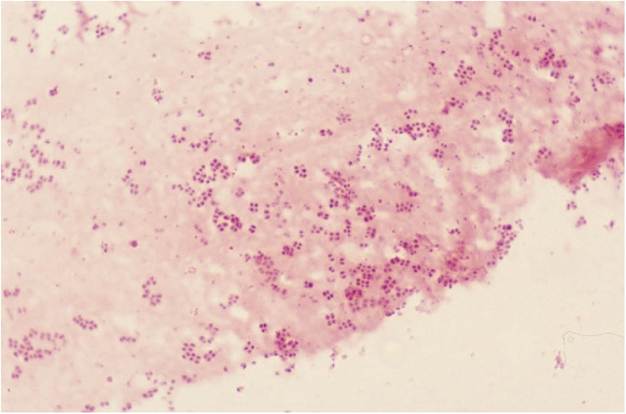
Figure 9-2
Modified Wright-Giemsa stain of staphylococci from a dog ear smear (magnification 1000?).
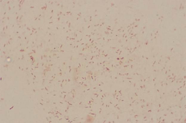
Figure 9-3
Gram's stain of Pseudomonas from a dog ear smear (magnification 1000?).
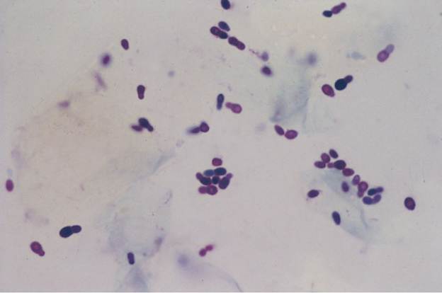
Figure 9-4
Modified Wright-Giemsa stain of Malassezia from a dog ear smear (magnification 1000?).
Many qualitative studies have been published concerning the microflora of the dog’s ear canal in health and disease. Tables 9-1 and 9-2 summarize the studies in the literature performed to evaluate qualitatively the microbial flora in dogs with normal ears and dogs with otitis externa.
Few studies have been performed to evaluate the microflora of the cat’s ear canal in health and disease. The most common bacteria recovered from otitis externa in cats’ ears are coagulase-positive staphylococci. Gram-negative bacteria such as Pseudomonas and Proteus spp. are only rarely recovered from cats with otitis externa.
Unfortunately, neither cytology nor culture are perfect diagnostic tests. In a recent study, cytology agreed with culture results only 68% of the time, but even more disturbing is that samples taken simultaneously from the same area of the ear canal gave different culture results 20% of the time.4
From a clinical standpoint, determining the significance of a yeast or bacterial population in animals with otitis externa is difficult because of the multifactorial origin of otitis externa. Many dogs and cats with otitis externa do not have increased numbers of microbes in the external ear canal. In addition, it has been theorized that microbes in the ear canal cannot proliferate unless inflammation or maceration occurs within the ear canal. Because of this, August considered microorganisms as perpetuating causes rather than primary or predisposing causes of otitis externa.3 In the semiquantitative cytologic evaluation study cited previously, the conclusion was that the pathogenic role of these organisms always depends on considerations other than only their numbers.3
Pathophysiology of Otitis Externa
Otitis externa has many etiologies. However, after the acute inflammatory stage has been initiated, there is a common pathway for the development of chronic otitis externa, regardless of the inciting cause. In the acute stage the ear canal becomes erythematous and swollen. Epidermal changes impede epidermal migration and decrease the self-cleansing function. The epidermis continues to thicken, and the sebaceous glands initially appear to become hyperplastic. These changes result in an increase in sebaceous secretion and desquamated cells, causing excessive wax production. As the otitis becomes more chronic, the apocrine glands begin to dilate and secrete. The addition of this low-lipid material decreases the concentration of the lipids secreted by the sebaceous glands. The lipid concentration of the cerumen decreases and the humidity in the ear increases.
Ultimately, the ear canal microclimate can be permanently altered. Stenosis of the canal and increased cerumen production can continually favor microbial overgrowth. Connective tissue proliferates in the dermis and subcutis, leading to fibrosis and additional thickening of the integument. The auditory cartilages may undergo calcification and possibly ossification, which further decrease the expansibility of the ear. All of these changes continue to cause further occlusion of the ear canal, which further impedes the normal cleansing function. It is likely that these altered ear canals will suffer from intermittently to constantly increased numbers of microbes, with attendant inflammatory changes, thus continuing the vicious cycle of chronic otitis.
More on the topic Otitis Externa:
- Otitis externa is a common malady, occurring in 15% to 20% of dogs and 5% to 7% of cats seen in veterinary practice.
- Most cases of acute otitis externa are pruritic.
- Factors that Predispose the Ear to Otitis Externa
- Perpetuating Factors and Treatment of Otitis Externa
- Careful examination of a clean, dry ear canal in a dog or cat with otitis externa may reveal many conditions that affect the ear canal.
- By definition, otitis externa represents a spectrum of inflammatory changes that occur to the external acoustic canal in response to any insult to the ear canal epithelium.
- Otitis Media
- Otitis media is a common disease process that often goes unrecognized in most veterinary practices.
- Chronic Inflammatory Otitis
- Primary Otitis Media in Cats
- Pathogenesis of Secondary Otitis Media
- Treatment of Otitis Media
- Environmental and Conformational Causes of Ceruminous Otitis
- Microbiology of Otitis Media
- Secondary Otitis Media in Dogs
- Otitis Media Prevents Healing
- The Use of Corticosteroids in Otitis Media
- Etiolog y of Ceruminous Otitis
- 14 Diagnosis and Treatment of Otitis Media
- 18 Otitis Interna and Vestibul ar Disease