TRAUMATIC INJURIES
Because the skin is in such an exposed position it regularly suffers from trauma. This may be due to physical injury, burns or chemicals. An example of the latter is tank cleaner inadvertently used as a teat dip.
Haematomas (Blood Blisters)
Haematomas most commonly occur on areas of the body where the skin covers bone. It is the pinching of the skin between bone and a hard object (e.g. a cubicle rail or narrow doorway) which leads to rupture of the blood vessel. The blood vessel continues to bleed, producing a large, soft, fluctuating swelling of blood under the skin. In areas where the skin covers muscle (e.g. on the ‘thick’ of the hind limb) there is a cushioning effect and haematomas are much less likely to occur. The common sites for haematomas are:
• the back, often caused by trauma from a cubicle rail (Plate 10.21)
• the ribs (from squeezing through doorways)
• the point of the shoulder
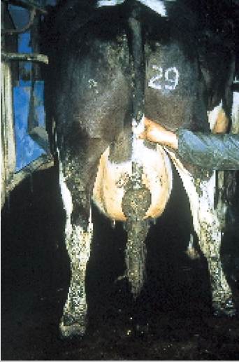
• the pelvis, especially beside the tail (Plate 10.22)
• over the hind leg, at the point of the stifle (Plate 10.23)
If left alone, the majority of haematomas will slowly disperse, although this may take several
Plate 10.21. Haematoma on the back, probably damage by a cubicle rail.
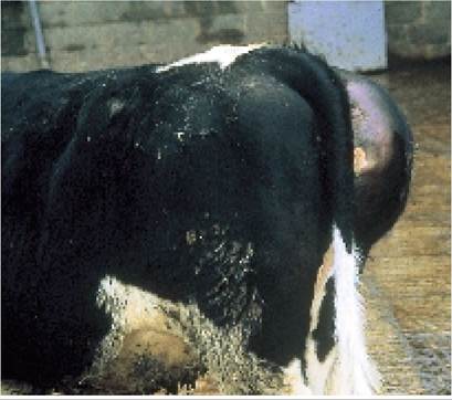
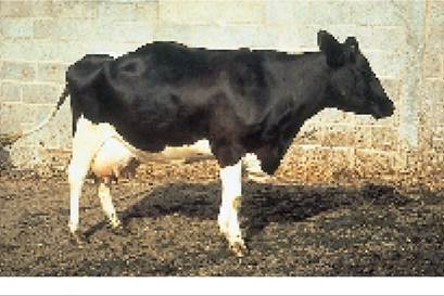
Plate 10.22. Haematoma beside the tail-head.
weeks. The problem with trying to lance and drain haematomas is that:
• The blood is not totally contained in a single ‘pocket’, so even if you open a large haematoma at one point, only a small area of it may drain.
• They may continue to bleed. This is especially the case if drainage is attempted only a few days after the haematoma has formed.
• Once opened, they can become infected and form an abscess.
In some cases this secondary infection can make the cow quite sick.For these reasons I would open a haematoma only if circumstances left no other option. The cow in Plate 10.22 is a good example. Blood from the haematoma had pushed through to the inside of the pelvis, putting considerable pressure on internal organs such as the rectum, vagina and urethra. This led to a cystitis and the cow was in considerable pain because she was unable to urinate.
In occasional herds ‘outbreaks’ of haematomas
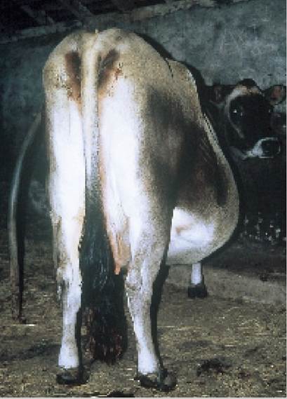
Plate 10.24. Flank hernia. This needs careful differentiation from a haematoma.
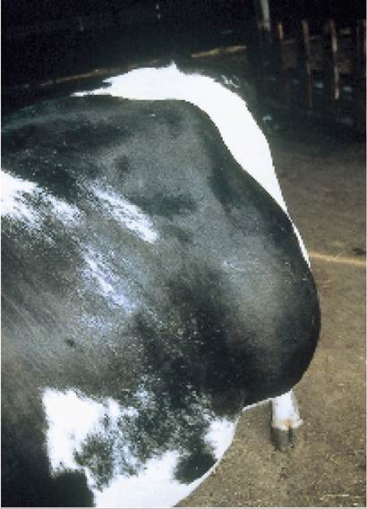
Plate 10.23. Haematoma on flank.
occur, where even normal everyday knocks will produce a haematoma, and large numbers of cows may be affected over a three or four week period. This is thought to be due to some factor interfering with the normal platelet aggregation process, thereby leading to increased bleeding, but no single factor has yet been implicated. I saw one herd where the syndrome appeared following a dietary upset which had led to severe ruminal acidosis. Others have suggested that it is a form of PPH (Chapter 13).
Haematomas should be carefully differentiated from abscesses and flank ruptures:
• Abscesses form slowly, gradually increase in size and are hard and often hot and painful. Haematomas are soft, fluctuating, painless swellings, which appear suddenly.
• Ruptures occur on the flank and may be differentiated (but not easily!) by feeling the tight edge of muscle which has torn to allow the intestine to pass through and lie under the skin. The Jersey cow in Plate 10.24 has a large rupture on her right flank.
Bursitis
A bursa is a small cushion of tissue acting as protection where skin covers bone and is often in an area where there is a considerable movement of the bone.
The best example is on the outside of the hock. If there is excessive and repeated pressure on a bursa (for example cows lying in hard cubicles with insufficient bedding), then fluid accumulates in the bursa to form a bursitis. The most common points of bursitis are over the hock (Plate 10.25) and on the neck (Plate 10.26). Neck bursitis is usually associated with cows continually pushing against a rail over the feed manger. Like haematomas, swollen bursae are best not drained; otherwise they may become infected. Ideally remove the cow from the continual trauma; for example take the cubicle housed cow into a straw yard, and the swelling will eventually disappear. Often this is not possible. Continual trauma will erode through the skin, resulting in an abscess (Plate 10.27).Abscesses
Abscesses can occur on any part of the body, although they are more common at points which project or can become damaged. Most abscesses are the result of infection penetrating the skin. The bacteria multiply and pus forms. The natural defence mechanism of the body is to stop the infection spreading, so it tries to encase the infection in a thick, fibrous capsule. This retains the infection in one place, but as the bacteria
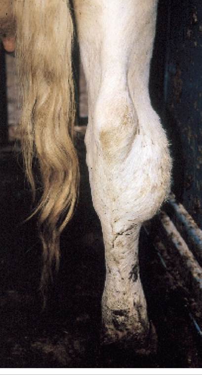
Plate 10.25.

Bursitis of the hock (‘cubicle hock'). At this stage the swelling contains only fluid and is not infected.
Plate 10.26. Bursitis of the neck, caused by the cow pushing against a feed rail
continue to multiply, pressure builds up within the abscess. Eventually the abscess capsule starts to weaken at one point - and this is where it eventually bursts.
The best treatment for an abscess is:
• Wait until it is close to bursting (you should be able to feel a soft point).
• Enlarge the hole from the ‘soft point’ downwards, thus allowing pus to drain from the bottom of the cavity.
• Wash out the abscess cavity daily. The easiest way of doing this is to insert a cold water hosepipe. Obviously do not use excessive pressure as this will be both painful and dangerous. If you do not flush an abscess regularly, there is a risk that it will heal with some infection left inside and then form again.
• Antibiotics by injection are not normally needed unless the cut through the skin was quite deep.
Although abscesses can occur at any point on the body, the most common sites are:
• the angle of the jaw (Plate 10.13)
• over the hock, often secondary to a bursitis
• secondary to neck bursitis, as in Plate 10.27. These are very difficult to treat, because they do not drain properly. Regular flushing with warm antiseptic solution is ideal, but even then expect it to take at least one to two months to heal
• in the muscle of the hind leg (Plate 10.28). These are known as popliteal abscesses. They originate from very deep within the muscle and are thought to be the result of infection from a previous foot or lower leg infection, draining via the lymphatic system. They must be left for a long time, probably one to two months, before drainage is attempted; otherwise the muscle incision needed will be so deep that the abscess will not drain properly.
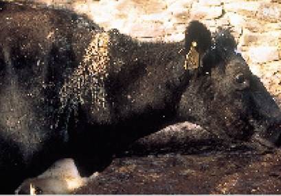
Plate 10.27. Neckabscessfollowing bursitis.
These do not drain well and so are very slow to heal. Regular flushing is essential.
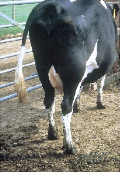
Plate 10.28. Popliteal abscess. Note the gross swelling of the right hind leg. The abscess must be left for one to two months before drainage; otherwise the incision through the muscle will be too deep.
Sterile Abscesses
Not all abscesses are caused by infection. Some result from inflammation caused by the injection of irritant liquids under the skin. The best example of this is the sterile abscess caused by the subcutaneous injection of a 40% calcium solution.
By no means all cows react to 40% calcium in this way, although it is more common if the whole bottle is injected into one site and the calcium is notdispersed (i.e. the site is not thoroughly massaged afterwards). Ideally, only 20% calcium solutions should be injected under the skin and the site should also be rubbed well after administration. Not only does the cow in Plate 10.29 have an unpleasant swelling on her side, but clearly the calcium was of no value to her, as it was not absorbed. Perhaps that is why she is having to be lifted on a hoist!
Cellulitis
If infection fails to localise, i.e. fails to form an abscess, then bacteria may spread through the tissues, usually just under the skin. This is known as cellulitis. Cellulitis occurs most commonly on the legs, often extending upwards from the hock and causing severe lameness (Plate 9.65). It also occurs around the face, where it may be part of the malignant oedema syndrome (Plate 10.14). Affected cows will have a high temperature and will need several days of treatment with injectable antibiotics.
Ingrowing Horns
Cows which have been badly dehorned, or where the horn has been damaged during growth, may develop an ingrowing horn. It is very easy to overlook the way the horn starts pushing into the skin beside the eye, especially when you see the cow every day and the change is slow. Plate 10.30 shows a typical example. The point of the horn can be removed using a wire or hacksaw. No anaesthetic is needed, because the ‘quick’ only comes about two-thirds of the way along the horn (see Plate 9.5).
Burns
Burns are relatively rare in cattle, although when they do occur they can cause quite severe damage. The cow in Plate 10.31 was one of a group of 30 dry cows, most of which were badly burnt when a large barn of straw adjacent to their shed caught fire and they were unable to escape. Badly damaged skin leads to shock due to pain and fluid loss. In the healing process the skin scars badly and contracts and often the hair never regrows.
If the teats or vulva are damaged this may produce permanent problems with calving and milking. Although the cow in Plate 10.31 was retained until after she calved, she proved impossible to milk and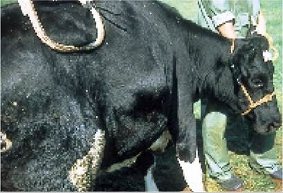
Plate 10.29. Sterile abscess caused by unabsorbed 40% calcium solution.
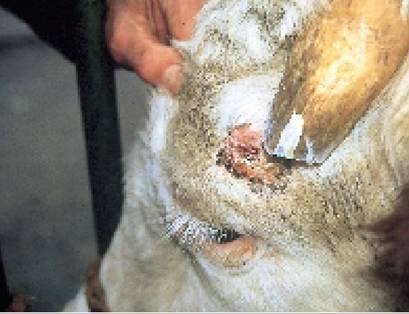
Plate 10.30. Ingrowing horn. This must be very painful, although unfortunately it is often unnoticed.
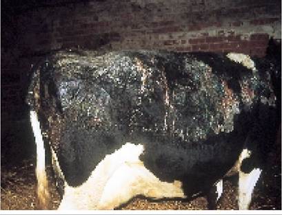
Plate 10.31. Severe burns caused by a straw fire in the adjoining building.
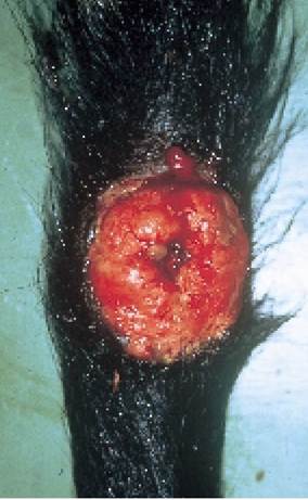
Plate 10.32. This discharging tail sinus is probably caused by a residual fragment of broken bone.
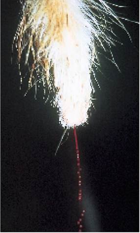
Plate 10.33. It can be very difficult to stop bleeding from the tip of the tail. This tail has been clipped ready for suturing.
had to be culled. It is interesting that even quite badly burnt cattle can be sent for emergency slaughter and in my opinion this is the best option, especially if they are insured.
For those animals which are retained, ensure that they are put onto a high-quality, high protein diet to restore the lost body protein. For younger calves, this often means putting them back onto a milk diet.
Tail Injuries
Tails commonly get broken. This may occur when cows are standing in overcrowded yards or cubicles; when the tail is caught in the diagonal of a gate; or unfortunately sometimes in association with rough handling. A simple break can be left untreated, but if an abscess or discharging sinus develops (Plate 10.32), or the tail tip is bleeding (Plate 10.33), intervention is needed. It can be quite difficult to stop bleeding from the tip of the tail by bandaging, probably because the blood vessel is held open by the bone of the tail. By far the best approach is to get your vet to give an epidural anaesthetic, remove the broken or infected fragment and suture across the tail tip.
Faecoliths are accumulations of faeces surrounding the tail. A typical example is shown in Plate 10.34. They should always be removed because the weight makes it uncomfortable for the cow
to lift her tail when passing dung, so the hind quarters and the udder get badly soiled. If left they gradually dry out, contract and erode into the tail. The bottom part of the tail may even drop off.
Tail marker tape (for parlour concentrate allocation etc.) can cause similar problems if it is left on for too long, or applied too tightly.
Plate 10.34. Afaecolith, an accumulation of dry faeces around the tail.
More on the topic TRAUMATIC INJURIES:
- Violence in the Mesolithic
- Trauma
- Aggression and violence are adaptive tools in the ancient evolutionary repertoire of humanity. So are cooperative and peaceful relations.
- Chapter 14 ROUTINE TASKS AND DEALING WITH POISONS
- 33 Intimate Partner and Sexual Violence
- Agrawal M.. Textbook of Pediatrics. 3rd ed. — CBS Publishers,2025. — 973 p., 2025