CHRONIC KIDNEY DISEASE
Chronic kidney disease (CKD) is defined as—(a) abnormalities of kidney structure or function for gt;3 months, with or without decreased GFR; or (b) GFR lt; 60 ml/min/ 1.73 m2 for gt;3 months without any structural or functional renal damage.
End-stage renal disease (ESRD) is the terminal phase of CKD, when life cannot be sustained without renal replacement therapy, e.g. dialysis or transplantation.
Severity of CKD is usually classified on the basis of GFR as per NKF-KDOQI classification (Table 21.16) or KDIGO classification. KDIGO classification also stage CKD in 5 grades (G1-5) similar to NKF classification, except sub-classifying Stage 3 into G3a (GFR 45-59) and G3b (GFR 30-44).
Etiology of CKD varies with age. While developmental renal anomalies and obstructive uropathies are common in under-five children, acquired lesions, e.g. glomerulonephritis, hemolytic uremic syndrome and hereditary nephropathies dominate in older children (Table 21.17).
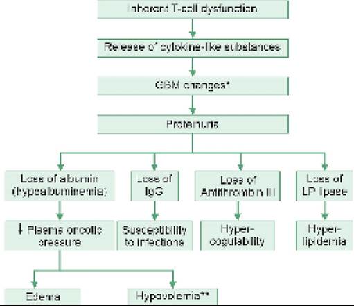
Pathogenesis: Kidney has a remarkable reserve to maintain its functions despite the loss of large number of nephrons, by compensatory hyperperfusion and hyperfiltration in remaining nephrons. However, such diversionary mechanisms further add to renal insult, probably by increasing hydrostatic pressure in surviving glomeruli, leading to proteinuria and progressive glomerular sclerosis (hyperfiltration injury).
*in ml/min/1.73 m2
NKF-KDOQI: National kidney foundation - Kidney disease outcomes quality initiative
Once a critical mass of nephrons in lost, either due to primary disease or hyperfiltration injury, progressive decrease in GFR ultimately lead to increasing severity of CKD and finally reaching to ESRD (Fig. 21.6). Proteinuria, hypertension and metabolic disturbances,
e.
g. hyperphosphatemia also contribute to progression of CKD.Terms chronic renal insufficiency, CKD and ESRD, are also used to signify GFR reduction, below 50%, 25% and 5% respectively.
Clinical manifestations of CKD appear insidiously with:
• Growth retardation due to poor nutrition, metabolic acidosis, anemia and reduced responsiveness to growth hormone. Puberty and skeletal growth is also delayed, with ultimate short stature.
• Edema due to fluid and salt retention.
• Hypertension due to renin-angiotensin mechanism and fluid retention.
• Normocytic normochromic anemia due to reduced erythropoietin production, uremic suppression of bone marrow and decreased RBC life span.
• Bleeding tendencies, e.g. GIT bleeding, due to thrombocytopenia (bone marrow depression) and defective platelet functions.
Fig. 21.6: Pathogenesis of chronic kidney disease.
• Renal osteodystrophy-a spectrum of bone diseases due to hyperphosphatemia (#936; excretion), hypocalcemia (secondary to hyperphosphatemia), compensatory hyperparathyroidism and vitamin D {1, 25 (OH)2 D3} deficiency. These cases present with refractory rickets, osteomalacia, osteitis fibrosa cystica and pathological fractures.
• Persistent metabolic acidosis, due to accumulation of non-volatile acids and loss of bicarbonates in urine.
• Neurological manifestations, e.g. altered behavior, ataxia, muscular weakness, neuropathy, often terminating in seizures and uremic coma, due to accumulation of toxic metabolites.
• Recurrent infections, due to granulocytopenia following bone marrow suppression.
• Skin itching due to uremic frost.
• Cardiac dysfunction due to cardiomyopathy, pericarditis or ventricular dysfunction may develop due to uremia, hypertension, fluid overload, dyselectrolytemia and vascular calcifications caused by mineral bone disease.
Laboratory evaluation of CKD includes:
• Complete urinalysis, including osmolality;
• Hematological: Hemoglobin, cell counts, platelet count;
• Renalfunction tests: BUN, S.
Creatinine and S. Uric acid;• Serum electrolytesquot; Na+, K+, Ca++, PO4-- and alkaline phosphate;
• Blood gas analysis, including serum bicarbonates.
• Skeletal survey for osteodystrophy and rickets
• Renal imaging, e.g. USG and renal scan, if required.
• Renal biopsy for definitive diagnosis
Periodic assessment of these parameters is necessary to assess progression of disease.
Management of CKD is largely conservative and aims to retard progression of disease and improve the quality of life till renal transplant is possible. Important components of management include:
• Nutrition: Diet in CKD should provide 100-120% of recommended caloric requirements as condensed feeds, including high biological value proteins (1-2 gm/kg/day), fats mainly in polyunsaturated forms (e.g. MCT oils) and water-soluble vitamins. Protein restriction is not advisable unless BUN exceeds 80 mg/dl, to avoid endogenous breakdown of proteins.
• Fluid and electrolyte management: Sodium and water restriction is not necessary except in cases with hypertension or terminal ESRD, when sodium intake should be restricted to lt;1 gm/day. Potassium restriction is necessary only in cases of documented hyperkalemia, which may also be controlled by oral alkalinizing agents and potassium exchange resins (PO Kayoxalate 1 gm/kg/day). Diet containing high potassium items should be avoided.
• Acidosis is controlled with oral NaHCO3 tablets (2-3 mEq/kg/day) with an aim to maintain serum bicarbonate levels gt;22 mEq/L. IV correction is advised if S. bicarbonate levels drop lt; 15 mEq/L.
• Renal osteodystrophy may be controlled by: (a) avoiding high-phosphorus foods, e.g. chocolates, dairy products, cola, etc., (b) use of phosphate-binders, e.g. calcium carbonate or Sevlamer hydrochloride (only in older children), (c) oral calcium supplements (0.5-2.0 gm/day), if hypocalcemia persists despite phosphate restriction, and (d) vitamin D supplements, preferably in active form (Rocaltrol 0.01-0.05 mg/kg/ day) along with cholecalciferol.
• Anemia is usually corrected by iron and folic acid supplements. Blood transfusions should be avoided unless hemoglobin falls lt;6 gm/dl, to prevent sensitization as these patients are potential candidates of renal transplant. Recombinant erythropoietin (SC/IV 150-300 U/kg/week) is recommended in cases with Hb lt;10 gm/dl to minimize the need for transfusion. However, EPO therapy may not work in presence of uncorrected iron deficiency and secondary hyperparathyroidism.
• Hypertension in early stages may be treated with diuretics, salt-restricted diet and ACE inhibitors, e.g. enalapril or Angiotensin receptor blockers, e.g. Losartan, though these agents should be avoided once GFR fall lt;60/ml/kg/min. Calcium channel antagonists, e.g. Nifedipine or amlodipine, are preferred in cases with GFR lt;30 ml/min. Severe hypertension may require additional therapy with clonidine or pazocin.
• Growth velocity, apart from good diet, may be optimized by recombinant growth hormone therapy (Sc 0.024 - 0.07 mg/kg/d), though high cost precludes its wider use.
• Recurrent infections need to be treated promptly, after modification of antibiotic doses as per creatinine levels. These children should also receive appropriate immunizations including those with HBV and all live vaccines in preparation for regular dialysis or renal transplant.
• Renal replacement therapy (RRT): Renal transplant and dialysis are two broad options for renal replacement therapy in ESRD and the choice of therapy and when to initiate it, depends on various clinical, biochemical, psychosocial and logistic factors. Renal transplant is always a preferred option for RRT, while Dialysis may be considered as a life-sustaining intervention till the transplant is possible.
Renal replacement therapy (RRT) should be considered when GFR falls lt;15 ml/min/1.73 m2 and strongly recommended when it drops lt;8 ml/min/1.73 m2, though the decision also depends on clinical status and presence of biochemical abnormalities.
Dialysis may be offered as continuous peritoneal dialysis (PD) or intermittent hemodialysis (HD), depending on the age and availability of procedure.
Continuous PD is usually preferred in ESRD children lt;5 years due to difficulties in getting satisfactory vascular access.• Continuous peritoneal dialysis can be provided at home through retention peritoneal catheters (e.g. Tenckhoff catheter), either as continuous ambulatory peritoneal dialysis (CAPD) or as intermittent cyclic peritoneal dialysis — manually or with the help of an automated device. Cycler-driven PD permits more freedom to the child who can continue to have normal activity in day-time and exchanges are performed automatically during sleep. Usually 4-6 cycles/day are necessary for satisfactory results. Peritonitis is a major complication, due to catheter-tunnel infections.
• Hemodialysis, usually twice or thrice a week, is more efficient option but requires adequate infrastructure, good vascular access and repeated hospital visits.
Renal transplantation (RT) is the treatment of choice in children with ESRD, using living-related donor (LRD-RT) or cadaveric kidneys (CAD-RT). Success rate depends on donor type, age of the recipient, and etiology of ESRD. Best results are obtained with LRD-RT, in children gt;2 years with non-hereditary disease.
Indications: Currently gt;60% of RT in children are performed for congenital or hereditary nephropathies. Table 21.18 provides reasonable criteria for selection of prospective candidates for RT.
Procedure: In brief, important steps in RT include:
• Evaluation of prospective recipient and donor to exclude serious infections and systemic diseases, and ABO and HLA typing.
• Recipient-donor matching for ABO and HLA type, as well as direct cross-matching between recipient serum and donor T and B cells, to detect pre-formed antibodies—potential cause of early graft rejection.
• Actual transplant surgery, with intensive intraoperative asepsis and fluid-electrolyte control.
• Post-transplant immunosuppressive therapy to prevent graft rejection and anti-infection prophylaxis against opportunistic infections.
Outcome: Overall success rate and survival among pediatric renal recipients is ~92-95%, higher among the LRD-RT than in CAD-RT. Opportunistic infections
during post-transplant immunosuppressive therapy is commonest cause of mortality in these cases, apart from other complications (Table 21.19). Renal disease may recur in the transplanted kidney.
21.11 TUBULAR DISORDERS
Renal tubules are primary sites for-(a) concentration and dilution of urine, (b) reabsorption or secretion of various solutes, (b) net acid secretion to maintain normal acidbase balance.
Renal tubular disorders includes primary defects of the generalized or specific tubular functions, without significant impairment in glomerular/interstitial function. Some important tubular disorders are discussed here.
21.11.1
More on the topic CHRONIC KIDNEY DISEASE:
- CHRONIC KIDNEY DISEASE
- Type 2 Diabetes
- Chronic Inflammatory Demyelinating Polyradiculoneuropathy
- Chronic Nephropathy
- ACUTE KIDNEY INJURY
- 19 Pancreatitis in a cat
- Crystalline Arthritis
- Extraskeletal Disorders
- Hypercalcemia
- Anemia of Chronic Renal Insufficiency