Diagnosis Based on Clinical Signs
Cattle Contrary to some diseases, bovine tuberculosis cannot be diagnosed with any degree of confidence based on its clinical signs. Mycobacterium bovis infection in animals causes a predominantly slowly progressive, often-asymptomatic disease in which clinical signs are usually not apparent until during the advanced stages of the disease (Fig.
9.1). The infection can remain subclinical for months (Thom et al.2004), and even years, until such time that infected organs are sufficiently affected and/or when the disease becomes generalized. The disease mostly affects the lungs and their draining lymph nodes, eventually causing ill-defined respiratory signs, emaciation, lethargy, lymphadenitis, fluctuating fever, anorexia, and ultimately death (Hines et al. 2006). This complex of clinical signs is well-known in parts of Africa, as in West Africa, BTB in Fulani cattle with coughing and loss of condition has for long been known as sondarou (Martrenchar et al. 1993). Advanced cases only occur in herds and in countries in which the disease has not been or is not controlled. Not all infected cattle develop the advanced or generalized disease, and more than 90% of cattle in a herd (Bonsu et al. 2000; Asiak et al. 2007; Menin et al. 2013) may remain asymptomatic, some of them for the duration of their life, with the infection only being detected at slaughter (Brush 1898). There is often no difference in the appearance and condition of cattle in herds with and without tuberculosis (Laval and Ameni 2004), and in Africa this may be the consequence of the presence of other chronic debilitating diseases such as contagious bovine pleuropneumonia, trypanosomosis, endoparasites, and malnutrition that often cause similar,
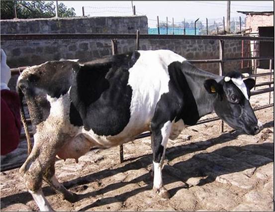
Fig. 9.1 A typically emaciated cow with advanced BTB in Addis Ababa, Ethiopia
non-specific clinical signs (Tschopp et al.
2010). When the infection localizes in organs other than the lungs, the clinical signs will vary accordingly. The inguinal lymph nodes in cows with mammary TB are usually enlarged, and the udder, depending on the nature of the reaction, may be visibly affected (Brush 1898).Small Stock, Pigs, and Camels Sheep, goats, and pigs have a similar distribution of lesions to those described for cattle, and they similarly develop clinical signs of marked, unexplained, chronic ill thrift, and occasionally diarrhea, with high mortality rates, and do not respond to symptomatic treatment (Hines et al. 2006; Bezos et al. 2012; Di Marco et al. 2012). In camels, a persistent high body temperature, cough, a profuse glairy discharge from the nostrils, pleurisy and/or peritonitis, cold and hard swelling of the superficial lymph nodes (Mason 1917), and gradual weight loss with inappetence occur (Kinne et al. 2006).
Wildlife Clinical signs in African wildlife vary substantially. Severe emaciation, lethargy, coughing, and death are characteristics in animals with advanced disease. These clinical signs are seen in greater kudus (Tragelaphus strepsiceros) (Keet et al.
2001), Kafue lechwe (Kobus leche kafuensis) (Gallagher et al. 1972), African buffaloes (Syncerus caffer), chacma baboons (Papio ursinus) (Keet et al. 2000), and lions (Panthera leo) (Keet et al. 1996). In African buffaloes, 75% or more of tuberculous buffaloes do not manifest clinical signs, and only 5% of BTB-positive animals usually have generalized lesions that would cause them to die within the course of a year (Jolles et al. 2005). In infected buffalo herds and particularly those with a high prevalence of BTB, coughing, that can be heard from a distance, is a distinct and regular feature (de Vos et al. 2001).
In greater kudus and bushbuck (Tragelaphus scriptus) with BTB, some have enlarged parotid and submandibular lymph nodes creating a large irregular swelling below the ear and at the angle of the jaw (Fig. 9.2) that justifies a presumptive diagnosis of BTB (Keet et al. 2001). Kudus are sometimes referred to as “roarers,” when lymph nodes containing tuberculous lesions press on parts of the respiratory tract and cause a roaring sound that can be heard at a distance when they breathe.
Fig. 9.2 Irregular swelling in the parotid and submandibular region of a kudu with BTB
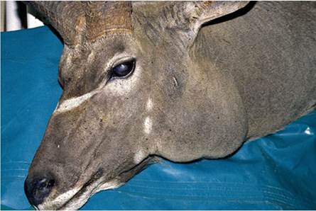
Fig. 9.3 A severely emaciated lion with BTB in the terminal stage of the disease
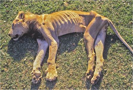
Lions, in addition to becoming severely emaciated (Fig. 9.3), also develop chronic arthritis with swelling of the elbow joints and large, ulcerated subcutaneous granulomas.
9.3
More on the topic Diagnosis Based on Clinical Signs:
- CLINICAL SIGNS AND TREATMENT
- Diagnosis
- Diagnosis
- TECHNICAL FACTORS OF NEEDLE ELECTROMYOGRAPHY
- Bovine Tuberculosis in Uganda
- Infections of the Genital Tract
- 1 Swallowing and regurgitation
- Hypertensive Disorders of Pregnancy: Preeclampsia/ Eclampsia
- 13 Endocrine Disorders of Pregnancy
- Chapter 9 Obstetric conditions