DISORDERS OF THE TEAT AND UDDER
Mastitis is defined as infection of the mammary gland. There are a few other conditions affecting the teat and udder which are worthy of note. Most of these are associated with physical damage, but a few are infectious.
Milking Machine Damage
The milking machine may damage teats by faulty pulsation, worn liners or improper use, for example removal of clusters while they are still under vacuum.
A certain amount of swelling (oedema) of the teat end (Plate 7.34) is a normal feature and is seen immediately the cluster is removed, but the teat sphincter should be smooth and not prolapsed. Plate 7.15 showed how milking machine damage could lead to eversion of the teat sphincter, often referred to as hyperkeratosis. There was also haemorrhage on the teat caused by excessive vacuum fluctuation. An advanced case of hyperkeratosis, associated with poor ACR function, was also shown in Plate 7.16. As a healthy sphincter is an important part of the defences against mastitis, teats with hyperkeratosis will be more prone to mastitis and will have increased cell counts.Blackspot
Blackspot is the term used to describe a particularly severe teat sphincter sore, usually consisting of a combination of an ulcerated area plus necrotic (dead) tissue, as in Plate 7.35. The ‘pull’ of the milking machine twice daily must retard healing and the damage to the teat canal obviously predisposes to mastitis.
Blackspot has no single cause. It usually starts with trauma, either by the milking machine or from crushing of the teat end. Secondary infection by the bacterium Fusobacterium necrophorum then
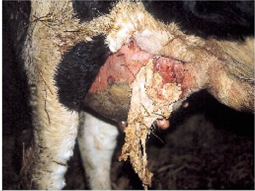
Plate 7.33. Tissue discharging following a gangrenous mastitis. This cow should be culled.
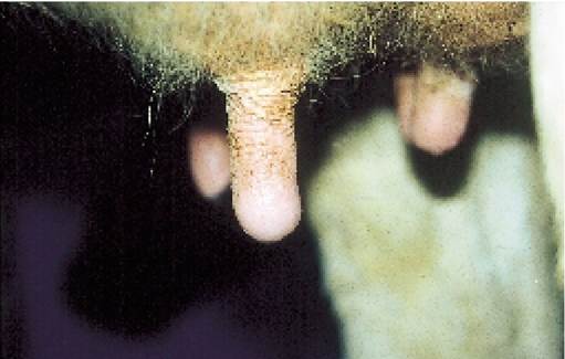
Plate 7.34.
Mild oedema or ballooning of the teat end, as in this cow, is a normal feature following removal of the milking machine.develops. Chilling by cold and wet weather exacerbates the condition.
Resting the teat, for example discontinuing milking for one or two weeks, or using a teat cannula (Plate 7.36) will help to improve healing, but both can lead to mastitis. If a teat cannula is used, make sure that a small quantity of antibiotic is deposited in the teat canal at the end of each milking for the four or five days following removal. The main risk of mastitis is after removing the cannula, presumably because the teat canal will have been badly stretched and its bacterial defence mechanisms will no longer be functioning. If you continue milking, remove the machine from that quarter as soon as possible. Teat dipping and antiseptic creams help to promote healing, as do ointments containing organic acids, which remove dead tissue. Some people have reported success using hypochlorite dips.
Cut Teats
Sometimes herds experience ‘outbreaks’ of deep gashes in their teats. The cut may run halfway round the teat or more; it is often at the lower end towards the sphincter and it may penetrate into the canal. A typical example is shown in Plate 7.37,
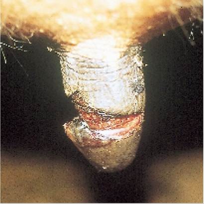
Plate 7.37. Atypical torn teat. The flap of skin is pulled downwards each time the unit is removed, making it very uncomfortable for the cow. Amputation of the flap promotes surprisingly rapid healing.
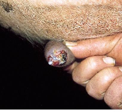
Plate 7.35. Blackspot is severe teat end damage caused by trauma and secondary bacterial infection.
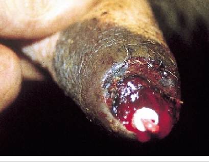
Plate 7.36. Ateat cannula used to allow milk to flow through a damaged teat end. The red plug can be removed to allow milk to flow.
and I know that the sight of this fills any herdsman with gloom. Teats which are split through the canal, as in Plate 7.38, are much more difficult to treat and often develop mastitis.
You will obviously need your vet to attend to the damage, but it may be worth looking at a few of the possible causes. The cut is most probably caused by the teat being stepped on, either by the cow itself, or by another cow. To try to prevent further cases you should look at possible overcrowding, cows being rushed about, poor cubicle design, insufficient cubicle numbers, inadequate dunging passage width, slippery floors and insufficient loafing areas leading to high stocking densities. It is also possible that you have a high proportion of older cows with pendulous udders, where the teats are more at risk.
Treatment is by suturing or by amputating the skin flap. Alternatively the wound can be taped over using a special adhesive plaster and aerosol spray (Plate 7.31). Although it is traditionally thought to be important to continue milking the teat because of the risk of mastitis, I think that mastitis is far more likely if a teat cannula is used (as in Plate 7.36). If the affected teat is simply left for one to two weeks, without being milked at all, this will promote much more rapid healing and most of the milk production from the teat will be
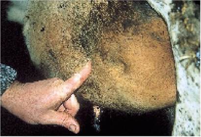
Plate 7.39. Udder oedema or ‘nature' is detected by pushing your finger into the udder. If a depression remains after removal, this indicates oedema.
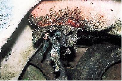
Plate 7.40. Necrotic dermatitis is seen in freshly calved heifers and is often a consequence of excessive oedema. The teat skin becomes very hard and dry.
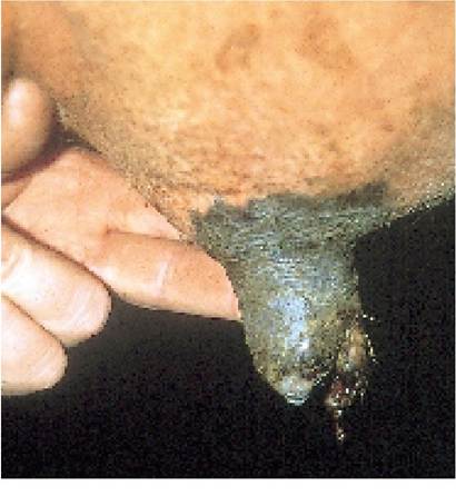
Plate 7.38. There is no specific treatment for a split at the teat end.
Infuse a small quantity of antibiotic daily and remove the milking machine quickly. The split may eventually heal.regained later. Make sure that milk from at least the first two milkings after resuming milking is discarded, as after two weeks the milk will have a very high cell count.
Udder Oedema and Necrotic Dermatitis
Oedema is the name given to fluid accumulating under the skin. It can be detected by pushing your finger into a swollen udder, then removing it. If a depression is left, the udder swelling is caused by oedema (Plate 7.39). At calving, udder oedema may be referred to as ‘nature’. It is caused by factors such as overfeeding, inadequate exercise and excessive salt or mineral intakes during the one to two weeks prior to calving. If severe, the oedema can restrict blood flow to the udder and teat skin to such an extent that the skin dies. This leads to the condition of necrotic dermatitis. Initially the affected skin will feel very hard and dry, and some areas may eventually fall off, leaving large sores.
Occasionally heifers may be so badly affected that they are almost impossible to milk. A typical example is shown in Plate 7.40. Sores may develop between the udder and legs (Plate 7.4l), or in older cows between the quarters at the front of the udder (Plate 7.42). These sores are very slow to heal. The best treatment is to wash with antiseptic solution, cleaning the area and removing all dead tissue, and then to dry and liberally apply an emollient such as glycerine. In the early stages, bathing the udder in a concentrated solution of warm Epsom salts may help to remove the oedema and restore blood flow.
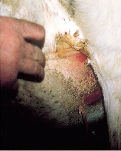
Plate 7.41. Sores may develop between the leg and udder, especially in freshly calved heifers.
Pseudocowpox
This is a paravaccinia virus and is probably the most common of the infectious teat lesions seen in cows. Typically it consists of irregular circular or horseshoe-shaped areas of small haemorrhagic spots, as in Plate 7.43.
There may be normal skin in the centre of the lesion. Sometimes the blister which precedes these changes may be noticed.As it is a virus infection, there is no specific treatment, although teat dip will help to prevent secondary bacterial infection and if mixed with an emollient it will promote healing. Hypochlorite also has a non-specific viral-killing action if in direct contact with the virus, although of course it
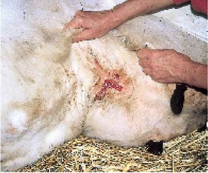
Plate 7.42. In older cows sores at the front of the udder are often first detected by their pungent smell!
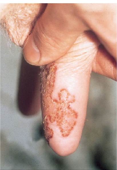
Plate 7.43. Pseudocowpox is a viral infection, seen as irregular circular shapes on the teat skin. It is not particularly painful. (Courtesy D. Weaver.)
is difficult to use with emollients (see page 199). If you have a severe outbreak you would be wise to milk the affected cows last to reduce the rate of spread of infection. Usually there are only a few cases in each herd, often in recently introduced heifers, because these have little or no immunity. Immunity to pseudocowpox is relatively short-lived anyway, and because of this some herds may experience waves of infection and disease every six to twelve months.
Gloves should be worn to prevent the development of milkers’ nodules, small warts on your hands and fingers which are caused by the virus. The virus is closely related to, if not identical with, the orf virus which causes scabs on the lips and nose of sheep and which can also affect man.
Bovine Herpes Mamillitis
This is another virus infection, fortunately much less common, because the disease is very severe. Large and very painful blisters develop on the teat and they may be so sore that milking is virtually impossible. When the blisters burst, a raw scabby area is exposed (Plate 7.44) and this may take two or three weeks to heal.
Cannulas may have to be used for milking. Heifers are most susceptible, and even the skin of the udder may be affected. At this stage it looks similar to a severe photosensitisation, but affecting only the teats and sometimes the skin of the udder. I have known freshly calved heifers to be so badly
Plate 7.44. Bovine herpes mamillitis is a much more severe viral infection which leads to teat blistering.
affected that they have had to be culled because they were impossible to milk. Luckily immunity is good, lasting four or five years, and herd outbreaks are relatively rare.
Teat dipping and separation of affected animals are the only useful control measures. Use an iodine teat dip, since iodine kills the virus. Disease is seen mainly from July to December, with September and October being the peak months, and most cases occur soon after calving. Even in a herd outbreak it is unusual for more than 10% of the cows to be affected, although the virus may persist in carrier cows for
many years before becoming reactivated.
Udder Impetigo
This is seen as small pustules, like weeping sores, over the skin at the back of the udder. It is caused by a staphylococcal infection and responds well to simple treatment with topical antibiotic or antiseptic cream. The teats are not affected.
Teat Warts
Warts are another virus infection and again it is heifers which are by far the worst affected, this time yearlings and in-calvers. Warts may appear as fleshy lumps (Plate 7.46) or they may be of the feathery type. Feathery warts (Plate 7.45) are the easiest to deal with because most of them can be quite easily pulled off and the teat dressed with an antiseptic cream or teat dip. With either type of wart you can ask your vet to send a specimen to a laboratory to have an autogenous vaccine prepared. The vaccine, which can be injected either into or under the skin, is probably only 30% effective, but sometimes heifers are so severely affected that any help is welcome. The virus is thought to be transmitted by flies, so attention to fly control (described on page 219) is important.
Body warts may also occur, with the head, neck (Plate 1o.15) and belly (Plate 7.46) being particularly badly affected. They occur mainly in cattle one to two years old and most cases spontaneously recover during the next summer at grazing. If the warts become so large that they ulcerate and develop a secondary bacterial infection, a vaccine can be prepared and this is much more effective than vaccines against teat warts.
Both types of warts can sometimes be prevented by mixing heifers with cows when they are younger, viz during their first grazing season.
Teat Chaps
This is the name given to cracks and splits in the teat skin. They become infected with bacteria which makes them sore, and of course they act as reservoirs of infection of the mastitis organisms Stap. aureus and Strep. dysgalactiae. The best treatment is teat dip or an ointment which has both antiseptic and emollient properties. Chaps occur
Plate 7.45. Feathery teat warts. These are caused by a virus infection and are most commonly seen in heifers.
particularly in the spring and autumn, when cows have to walk through muddy gateways and when there are cold winds. Teat skin does not have the sebaceous glands found elsewhere in the body. This means that when dry, the normal pliable and elastic properties of the skin are soon lost, its keratin layer cracks, and chaps soon form. This is one reason why pre milking teat washing is being discontinued, especially in herds which do not dry teats afterwards.
Milk Let-down Failure
Milk let-down, that is the expulsion of milk from the glands where it is produced (Figure 7.1) into the teat, is stimulated by the hormone oxytocin.
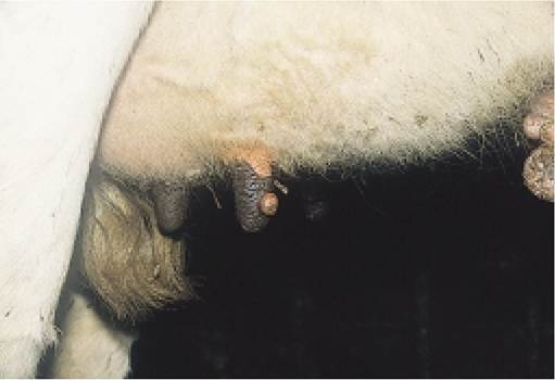
Plate 7.46. ‘Fleshy' teat warts are also caused by a virus.
In some cows the normal activities of entering the parlour, feeding and udder preparation do not seem
sufficient to stimulate oxytocin release, and virtually no milk is given. This can be particularly true with heifers which are apprehensive or nervous, because the hormone adrenalin acts as an antagonist to oxy-
tocin. The problem can be approached in two ways. First, careful and gentle handling may overcome the
heifer’s fears and, second, injections of oxytocin can be given two to three minutes before the milking
machine is applied. In practice you would probably use both methods. When you have established the
dose of oxytocin required to produce let-down, try slowly decreasing it over a few days until the heifer’s
own behavioural reactions take over.
Sometimes cows which are being suckled, or are suckling others, stop letting down their milk. Provided you can identify the culprit, most cases can be controlled with an anti-suckling nose plate (as in
Plate 7.47), which covers the cow’s mouth and prevents her getting hold of a teat. It is said that group-housed calves which are allowed to suckle each other during rearing are more likely to suckle as adults. Ideally calves should be penned individually until well after weaning.
Blind Quarters
This is a condition seen primarily in heifers, and would not be noticed until the first milking. The udder appears normal, but no milk can be drawn from the teat. I have experienced three separate categories of this condition. The first, and by far the most common, is the presence of a membrane across the top of the teat, producing a permanent barrier between the teat cistern and the gland cistern (see Figure 7.1). The teat feels normal but it does not fill with milk during let-down. The teat is anaesthetised and a long cannula (often called a teat siphon) is inserted through the teat sphincter. Often the cannula can be forced through the membrane to allow milk to flow. Making a series of holes in this way can occasionally resolve the blockage, but many eventually heal over again.
The second cause of a blind quarter is a blockage at the teat sphincter. The teat feels full of milk, but it cannot be drawn out. This is the easiest condition to deal with. With the teat anaesthetised, a small knife (called MacClean’s knife) with a disc blade
just below the guide tip is forced up through the sphincter as shown in Plate 7.48. It is then rotated
through 180° and pulled back out again. This produces two transverse cuts, and once milk starts to flow,
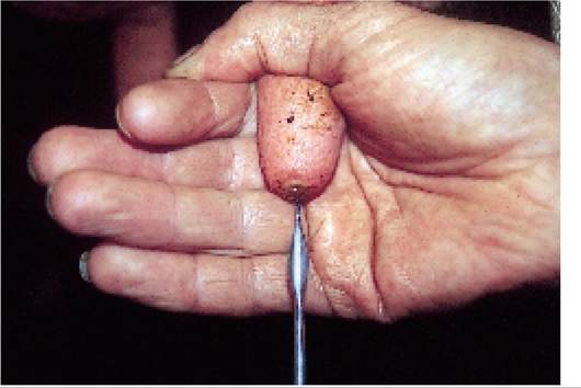
Plate 7.48. A MacClean’s teat knife, used to dilate the teat canal of a ‘tight’ (slow) milker and also to remove teat ‘peas’.
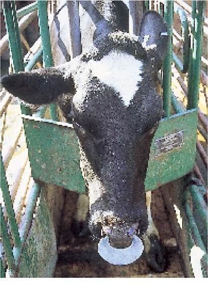
Plate 7.47. An anti-suckling nose plate. Some plates also have protruding spikes, which discourage the cow doing the suckling.
it usually continues very successfully, although it may be best to infuse intramammary antibiotic into the teat end after each milking for the first few days as a mastitis preventive. The same procedure can also be used to dilate the teats of cows or heifers which are very slow milkers, provided the sphincter is normal. If the slow milking arises from a crushed teat or some other abnormality, however, I have not found the knife particularly successful.
The third cause of a blind quarter is summer mastitis. The heifer will already have had the infection, quite possibly unnoticed, and may well have recovered without treatment, but the teat is left permanently damaged. It feels as if there is a thick fibrous
core running up through the teat cistern. This is easily detected by rolling the teat between your finger and thumb and comparing the affected teat with a normal one. There is no treatment.
Blood in Milk
It is not uncommon for cows to calve down with blood in their milk, and I have always felt that it is more common in animals which have very tight oedematous udders or sometimes following a difficult calving when the udder may have been bruised by the cow’s own leg movements. Sometimes the blood has formed clots and then the diagnosis is easy. At other times it is mixed with colostrum, and it may be very difficult to decide if there is an acute mastitis present. Looking for a raised temperature, heat and pain from the quarter and general signs of health should distinguish between the two conditions, but if you are in any doubt I would strongly recommend that you infuse a tube of antibiotic. I know of no drugs which are consistently effective against blood in milk, and the only action is not to milk the quarter or to relieve it only lightly so that the back pressure from the milk stops the blood flow. There is some evidence that cows will develop a ‘light’ quarter, that is they will not milk as well, after they have had blood in their milk.
Pea in Teat
Sometimes milk flow from the teat is obstructed by a small lump which floats around in the teat cistern but acts like a valve as soon as milk is drawn from the sphincter. This is called a ‘pea’. Examples are shown in Plate 7.49. The red-coloured ‘peas’ are very like blood clots, which suggests that they could be a consequence of blood in milk, with milk salts, fat and udder cells then adhering to the blood clot in the more mature (cream-coloured) cases. ‘Peas’ can occur at any stage of lactation, but are most commonly seen during the first three months after calving, when the cow is at peak yield.
Small peas may be squeezed out manually, perhaps by first crushing them inside the teat, using your finger and thumb. However, in the
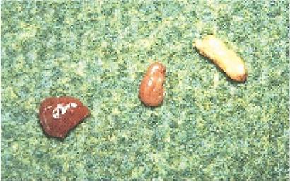
Plate 7.49. Examples of teat ‘peas'. Some are quite soft, like a blood clot, which suggests this could be their origin.
majority of cases the sphincter has to be dilated with a MacClean’s knife (Plate 7.48) to facilitate removal. Alternatively, use a pair of special forceps inserted through the canal to crush the pea and then pull it out in pieces. Sometimes the pea is attached to a membrane growing out from the wall of the teat cistern, or it is the membrane itself which is causing the blockage. Although milk flows easily through a cannula, as soon as the teat is drawn by hand the membrane obstructs the teat streak canal. I have never found any successful way of treating such cases.
Chapter 8
More on the topic DISORDERS OF THE TEAT AND UDDER:
- TEAT AND UDDER DEFENCES AGAINST MASTITIS
- Chapter 7 MASTITIS AND CONDITIONS OF THE TEAT AND UDDER
- POST MILKING TEAT DISINFECTION
- P Keerthi Kundana, Mona Gajre, Alpana Kondekar, Mukesh AgrawalNeurological disorders account for ~15-20% of hospitalizations, which may be divided into three major categories: (a) central nervous system disorders, involving brain and spinal cord, (b) neuromuscular disorders involving peripheral nerves and muscles, and rare disorders of autonomic nervous system.
- Mastitis continues to be a major cause of economic loss to the national dairy herd and I suspect that, combined with teat injuries, it is one of the greatest aggravations to the herdsman.
- Rheumatic disorders (connective tissue disorders or collagen vascular disorders') is a collective term to denote a large group of conditions with variable manifestations but two common characteristics: (a) acute or chronic inflammation of multiple target organs, specially musculoskeletal system and vasculitis, and (b) an underlying abnormal immune response, e.g. autoimmune etiology.
- DISORDERS OF DERMIS DISORDERS OF SUBCUTANEOUS FA
- MOVEMENT DISORDERS
- IMMUNODEFICIENCY DISORDERS
- HEMORRHAGIC DISORDERS
- SYSTEMIC CONNECTIVE TISSUE DISORDERS
- SPINAL CORD DISORDERS
- Vesiculobullous disorders
- ANXIETY DISORDERS
- NEURQDEGENERATiVE DISORDERS
- DISORDERS OF LIPID METABOLISM
- LEUKOCYTE DISORDERS
- AUTISTICSPECTRUM DISORDERS
- Esophageal Motor Disorders