TEAT AND UDDER DEFENCES AGAINST MASTITIS
Before discussing practical aspects of mastitis control, the natural defence mechanisms of the teat and udder will be examined. This will enable the reader to appreciate more fully the reason why he is carrying out certain procedures in the milking parlour.
The section is subdivided into teat defences and udder defences.Teat Defences
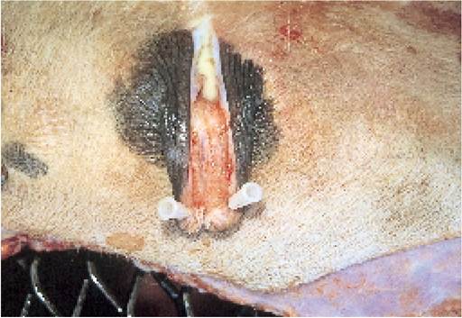
Plate 7.1. Section through a teat showing the interlocking folds of keratinised epithelium of the sphincter.
The teat has a number of ingenious defence mechanisms aimed at preventing the entry of bacteria and reducing the chances of mastitis (Figure 7.1). The outer layer of teat skin, called stratified squamous epithelium, has a lining of dead cells, all impregnated with a hard, inert material called keratin, and this does not easily support bacterial growth. Only when the teat is cracked or chapped can large numbers of bacteria grow on the surface of teat skin. Secondly the physical tightness of the teat sphincter muscle keeps the streak canal firmly closed and this helps to prevent bacterial entry. Third, the streak canal is also lined by keratinised epithelium (Plate 7.1), the superficial dead cells of which attract, and trap bacteria which may be invading. When milk flows out, both bacteria and dead cells are flushed away from the udder. Perhaps most interesting of all, the epithelial lining around the teat end and through the streak canal contains lipids and proteins which have specific antibacterial activity. These protein molecules are even positively charged so that they can attract negatively charged bacteria towards them before damaging their membrane and destroying them. In addition, the lipid surface lining of the teat canal acts as an extra ‘seal’. At the end of milking the teat sphincter contracts and this pushes the opposing surfaces of the teat canal together, thereby either expelling any residual milk, or at least breaking the milk column up into small ‘lakes’.
There is then no longer a solid column of milk through which bacteria can track back into the teat, ie any ‘wick’ effect has been eliminated. These small residual lakes of milk can be removed by foremilking before the clusters are attached at the next milking. If the teat canal has been damaged the lipid seal may not be complete, and any serum oozing from the cracked teat canal would act as a nutrient for invading bacteria.The inside of the teat cistern is lined with a similar type of epithelium (but it is not keratinised) and this provides a further defence against certain types of bacteria, although others may be able to establish colonies in this area.
Teat closure and mastitis
At the end of milking the small sphincter muscle around the tip of the teat canal (see Figure 7.1 and Plate 7.1) contracts, thus closing the teat. This considerably reduces the chances of bacteria entering the teat between milkings. Under the stimulation of milk let-down (provided by either the calf or the milker), engorgement of erectile tissue around the base of the teat makes it become turgid and holds it open. It then fills with milk and the teat sphincter relaxes to allow milk to flow.
The pressure required to force bacteria back up through the teat canal is therefore much less during milking than between milkings. Approximate pressure figures required are:
-15 kPa before milking
- 5 kPa during milking
- 3 kPa at the end of milking
-15 kPa 30-40 minutes after milking, when the sphincter has closed again
(kPA = kilopascal, a measure of vacuum. One inch of mercury (Hg) is equivalent to 3.33 kPa)

Time after milking
--------------- >
Figure 7.3. The importance of teat sphincter closure and E. coli mastitis. Thirty-five per cent of the teats which were exposed to a culture of E. coli in the first ten minutes after milking developed mastitis, whereas this fell to only 5 per cent when the culture was applied one hour before the next milking.
Figure 7.3 shows the effects of applying a culture of E. coli to the teat end. If applied 10 minutes after the end of milking, 35% of quarters developed mastitis, whereas this fell to only 5% of quarters if the culture was applied one hour before the next milking. The practical implications of this are that cows should be kept standing, in a clean environment, for at least 30 minutes after milking. If they walk through a dirty cubicle passage and then lie down with their feet against their udder while the teat ends are still open, the risk of mastitis can be enormous.
The increasing openness of the teat sphincter, leading to increased susceptibility to mastitis, was referred to on page 175. When called to treat severely ill cases of down-calving mastitis, often the affected cow is a very easy milker with a very open teat end. Milk may flow out easily, but unfortunately bacteria can get in equally as easily! We must therefore milk and manage our cows in such a way as to minimise this ever-increasing risk.
In summary, the defence mechanisms of the teat to counteract bacterial invasion include
• Keratinised squamous epithelium is a hostile environment for bacterial multiplication.
• Contraction of the teat sphincter between milkings closes the canal.
• An inner lipid layer completes the seal.
• Specific lipids and proteins have antibacterial properties.
• The surface layers of keratin which adhere to invading bacteria are flushed away at the next milking.
Udder Defences
Even when bacteria have penetrated the many defences of the teat, it is by no means certain that they will become established in the udder to cause mastitis. Probably the most effective means of eliminating recent invaders is the milk flow itself: the majority of bacteria entering the udder are simply flushed back out again. For those which remain there is a range of very effective defence mechanisms within the udder to deal with them. These mechanisms are:
• intrinsic defences in milk
• macrophages in milk
• neutrophils from the blood
Intrinsic defences
Milk contains a range of bacterial inhibiting systems.
For example, lactoferrin is present in the dry cow and prevents E. coli multiplication; lactoperoxidase is probably important in the control of Streptococcus uberis; immunoglobulins in milk coat the surface of bacteria and render them more susceptible to phagocytosis by macrophages and neutrophils.Macrophages
Macrophages are large cells present in the milk which are capable of engulfing and destroying bacteria. This is known as the process of phagocytosis and was described in Chapter 1, Figure 1.3. Acow with a high cell count has an increased number of macrophages and neutrophils in her milk. Although macrophages assist in the control of infection and provide the primary line of defence, they are not the major ‘attack force’. This consists of the neutrophils, or to give them their full name, polymorphonuclear cells, PMNs.
Neutrophils
Neutrophils are white cells which can pass from the blood into the milk in huge numbers in response to an alarm signal sent out by the macrophages. An analogy could be made between the ‘bobby on the beat’ (macrophages) and a ‘rapid reaction force’ (neutrophils).
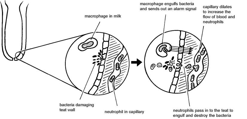
Figure 7.4. The response of the teat to bacterial invasion. This reaction can also take place in the udder cistern and ducts.
The ‘alarm signal’ consists of a combination of waste products, produced as the macrophages destroy bacteria, plus toxins released by the multiplying bacteria themselves. E. coli is an especially active producer of toxins. This is why in some cows we see an extreme udder response to E. coli infections. The sequence of events following release of the alarm is shown in Figure 7.4 and is as follows:
1.
2.
3.
Mammary blood vessels including capillaries in the teat wall dilate to carry more blood (and therefore neutrophils) to the udder. This is why a mastitic cow may have a hard, hot, swollen and painful quarter (Plate 7.2).
The cells lining both the teat wall and the capillaries move apart, so that neutrophils can pass through into the milk. This also allows leakage of serum, which is why in many cases of E. coli mastitis the ‘milk’ appears brown-coloured, like serum.Once into the milk the neutrophils rapidly locate and then destroy the invading bacteria. This produces more ‘signals’ and further amplifies the alarm. After a severe bacterial
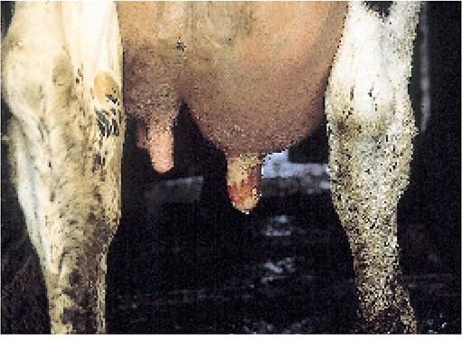
Plate 7.2. Swollen quarter, typical of mastitis. The teat has been damaged.
invasion of the udder the neutrophil response can be so effective that almost all the white cells are drained out of circulation and blood counts may fall to almost zero! At the same time the cell count of milk may rise from a background level of around 100,000 (105) per millilitre to as high as 100 million (108) per millilitre within a few hours. In this case the majority of the cells would be neutrophils - and if milk from just one mastitic quarter were to enter the bulk tank, the bulk milk cell count would rise dramatically!
Although the whole process can be in operation in as little as four hours from the entry of E. coli, cows vary enormously in the rate at which their neutrophils can mount a counter-attack, and also in the ability of their neutophils to kill bacteria. In one experiment, some cows were able to destroy 98% of the E. coli infused into a quarter in as little as six hours, whereas other cows destroyed only 80%. This variation in activity, which is probably genetic, is seen at any age and at all stages of lactation, so heifer calves could be blood sampled to assess their ability to withstand mastitis infection. Research is even being carried out on bulls by taking a sample of their blood and monitoring the response of their neutrophils to chemotaxin and E. coli attack. In this way it may be possible one day to predict those bulls that will produce daughters with a rapid neutrophil response to E.
coli; that is those which are able to easily counteract E. coli mastitis.Stage of lactation and response to infection
Numerous surveys have shown that the highest incidence of mastitis occurs around the time of calving and this is particularly true for coliform infections. In some ways this finding is rather surprising, because the freshly calved cow has high levels of antibody in her colostrum and this might make you think she would be more resistant, rather than more susceptible. There seems to be something about the freshly calved cow which reduces her ability to mount a good response to infection. This is demonstrated in Figure 7.5. Figure 7.5a shows a good response in a mid lactation cow. There is a rapid rise in neutrophils and the E. coli are quite quickly eliminated from the udder. The herdsman sees this clinically as a cow with a hard, hot and swollen quarter, probably producing a brownish, watery secretion. The cow may well have a raised temperature and be off-colour, but if a milk sample is taken, it could well be sterile: the inflammatory response mounted by the cow was so effective that all the bacteria were eliminated in six to eight hours and the herdsman is simply seeing the residual damage caused by the E. coli.
Contrast this with the situation in Figure 7.5b, which is a freshly calved cow. For some reason she was unable to mount a good neutrophil response. The E. coli continue to multiply unchecked and huge numbers are present in the udder. (If this quarter was milked into the tank, bulk milk TBC would soar!) There is no swelling or hardness in the quarter because the cow has been unable to mount an inflammatory response, and initially there are probably very few changes in the milk, but she will be very sick, probably scouring, unable to rise and with sunken eyes. These changes are produced by the release of endotoxin from the bacterial cell walls. Even the use of antibiotics will have a limited effect: the cow needs treatment for generalised shock, since many of her body organs will be affected by endotoxin.
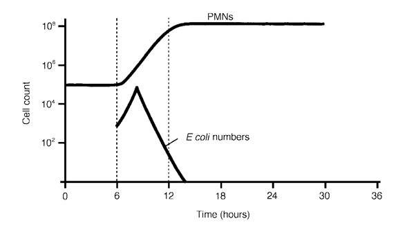
Figure 7.5a. Good neutrophil response in a mid lactation cow can lead to rapid elimination of E. coli.
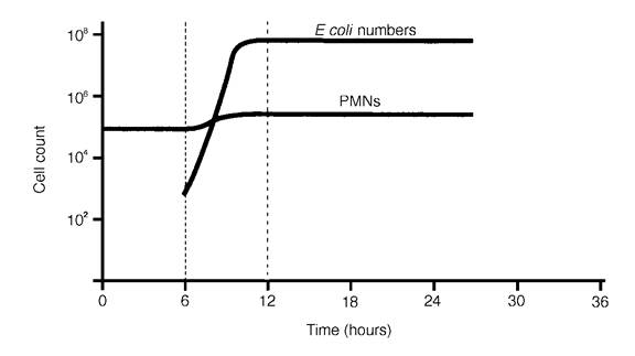
Figure 7.5b. The poor cellular response seen especially in some freshly calved cows allows E. coli to multiply to very high numbers in the udder (compare this with the good response shown in Figure 7.5a). Provided the cow survives, bacterial numbers may remain high for several days.
From Hill, A. W. (1981) Res Vet Sci, 31, p. 107.
The major factors which influence the effectiveness of the teat and udder at controlling invasion by mastitis bacteria are:
• the quality of teat skin, especially at the teat end: poor-quality cracked skin predisposes to bacterial growth
• the openness of the teat canal
• the speed at which neutrophils can pass from the blood into the udder
• the ability of those neutrophils to engulf the bacteria in milk
• the nature of the invading organism: does it produce toxins (E. coli) or does it have adhesive properties (Staph. aureus)?
• stage of lactation: the freshly calved cow is particularly bad at mounting a response against E. coli
Response to Staphylococcal and Streptococcal Mastitis
Staphylococci and streptococci invade the deeper parts of the gland and evoke a similar neutrophil response, although much less intense. These organisms differ from E. coli in that they have adhesive properties. Because they stick onto the inside of the udder tissue, they are much more difficult to eliminate, even though macrophages and neutrophils kill some of them.
It is important to remember the following:
• Staphylococci and some streptococci produce persistent infections. Persistently infected cows then act as a reservoir of infection for other cows.
• E. coli does not have adhesive properties and does not persist in the udder. Other cows are therefore not reservoirs of infection for E. coli, its major reservoir being the environment.
With staphylococcal and streptococcal infections the smaller udder ducts may become blocked with clumps of bacteria, neutrophils and general debris (Figure 7.1). By this stage the alveoli will no longer be producing any milk. The blockage may become almost permanent, and it will then be difficult for antibiotics to penetrate the foci of bacteria trapped inside. Some bacteria will periodically leak out during the course of the lactation, however, to evoke an inflammatory response in adjacent alveoli. This is seen clinically as a recurrent case of mastitis, but even if no clots are evident the cow will be intermittently shedding bacteria in her milk and will therefore be a danger to others. A cow with a chronic Staph. aureus infection often has a hard and swollen quarter, as shown in Plate 7.3.
With the virtual elimination of Staph. aureus from many farms,
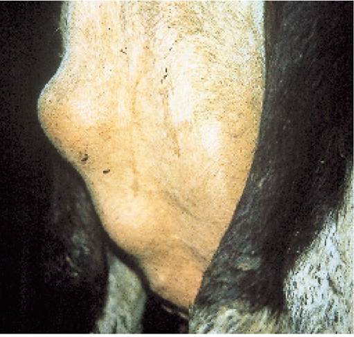
Strep. uberis is now becoming a more Plate 7.3. Swollen quarter typical of chronic Staph. aureus. This common cause of chronic recurrent cow consistently showed a cell count of 3 million. mastitis. Although it probably starts as an environmental organism, chronic Strep. uberis infections should undoubtedly be classified in the ‘contagious’ category. Natural antibody defence mechanisms within the udder are relatively poor at eliminating Strep. uberis, and many strains of the organism appear to be able to resist phagocytosis by macrophages and neutrophils. Why penicillin appears so ineffective against such strains remains unknown, because all strains are sensitive to penicillin on the plate test (treatment is discussed in detail in a later section).
More on the topic TEAT AND UDDER DEFENCES AGAINST MASTITIS:
- Chapter 7 MASTITIS AND CONDITIONS OF THE TEAT AND UDDER
- DISORDERS OF THE TEAT AND UDDER
- Mastitis continues to be a major cause of economic loss to the national dairy herd and I suspect that, combined with teat injuries, it is one of the greatest aggravations to the herdsman.
- POST MILKING TEAT DISINFECTION
- THE MILKING ROUTINE AND MASTITIS CONTROL
- THE ENVIRONMENT AND MASTITIS
- SUMMER MASTITIS
- MASTITIS RECORDS AND TARGETS
- THE CONTROL OF MASTITIS
- Physical and Chemical Defences
- WHAT IS MASTITIS?
- TREATMENT OF MASTITIS
- 8 Compliance and Non-Compliance – Defences