Rotavirus Infection: Epizootic Diarrhea of Infant Mice
Epizootic diarrhea of infant mice (EDIM) virus belongs to the family Reoviridae, genus Rotavirus. The rotavirus A group encompasses viruses of humans, nonhuman primates, cattle, sheep, horses, pigs, dogs, cats, turkeys, chickens, and rabbits that are closely related genetically and antigenically.
Each of these viruses has relative host specificity, but interspecies infection can be shown experimentally with high doses of inocula. They share a common propensity to cause enteritis and diarrhea in infants. EDIM has been studied as an animal model for rotaviral infections in other species. A number of group A rotaviruses have been isolated from mice, of which EDIM virus is a single strain. However, the term "EDIM” has been generally accepted as the inclusive name for intestinal rotavirus infection in mice and its agent.Epizootiology and Pathogenesis
EDIM virus is highly contagious and prevalent in both laboratory and wild mice. Disease manifestations are relatively rare once infection is enzootic, due to protection of clinically susceptible infant mice by maternal IgA. Typically, clinical signs of EDIM occur in naive breeding populations, but once infection is enzootic within the colony, EDIM disease is no longer apparent, although EDIM virus remains. Rotaviruses are shed copiously in feces, and transmission is by the orofecal route. Clinical disease ranges from inapparent to severe, depending primarily upon age. All ages of mice are susceptible to infection; however, disease is limited to mice less than 2 weeks of age. Virus selectively infects terminally differentiated enterocytes of villi and surface mucosa of the small and large intestine, respectively. These cells are most plentiful and widespread in the neonatal bowel and diminish in number, distribution, and degree of terminal differentiation as mucosal proliferative kinetics accelerate with acquisition of intestinal microflora.
Viremia is detectable during peak virus replication, but virus replication is restricted to intestinal mucosa. All ages of mice are susceptible to productive infection, but the target cell population in adults is limited. Thus, functional disturbances tend not to be noted in older mice. Regardless of age at infection, recovery from diarrhea occurs at 14-17 days of age, with complete recovery from infection. Susceptibility to infection and disease may have a genetic basis, with BALB/c mice relatively susceptible, and B6 mice resistant. Infection of SCID mice follows the same age- related pattern of disease as immunocompetent mice. B cells, CD4 T cells, and CD8 T cells all contribute to resolution of infection, with persistent shedding of virus in B-cell-deficient and SCID mice.During the first few days of infection, there is fluid accumulation and dilation of the small intestine. Diarrhea is induced by a combination of factors, including apoptosis and loss of absorptive epithelium, replacement of lost cells with immature nonabsorptive cells, altered carbohydrate absorption, osmotic effects related to luminal carbohydrate and bacterial fermentation, and active secretion of fluid and electrolytes. The secretory
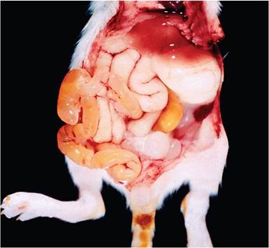
FIG. 1.42. Infant mouse with epizootic diarrhea of infant mice (EDIM), caused by rotavirus. Note the full stomach, the flaccid and dilated small intestine, and pasting of the tail base with soft feces.
stimulus appears to be mediated by an enterotoxic effect of the viral nonstructural protein 4, which can be evoked with inactivated virus or recombinant protein, and activation of the enteric nervous system. Overgrowth of E. coli, and its atypical abundance in the upper small intestine, accompany the malabsorptive effects of the virus.
Pathology
Infection is often silent, but clinically affected mice can be runted and potbellied, with loose, mustard-colored feces staining the perineum.
Steatorrhea with oily hair may also be apparent. The bowel is flaccid and distended with fluid and gas (Fig. 1.42), but mice continue to suckle. Some deaths may occur due to obstipation caused by fecal caking around the anus. In infant mice, EDIM virus causes hydropic change and vacuolation of terminally differentiated enterocytes at the tips of villi (Fig. 1.43) and large intestinal surface mucosa. Some nuclei may be pyknotic. Acidophilic intracytoplasmic inclusions have been described but are not diagnostic. In addition, the lamina propria may be edematous and lymphatics dilated, although inflammation is minimal. These changes are difficult to discern under the best of circumstances and are not apparent in mice older than 14 days of age. Remarkably, mice may manifest significant diarrhea with minimal microscopic lesions. Infected infant mice often have severe stress-related thymic necrosis.Diagnosis
EDIM can be diagnosed presumptively on the basis of age, clinical signs, and lesions. Differential diagnoses include enterotropic MHV, MAdV-2, reovirus, salmonellosis, Tyzzer's disease, and Clostridial enteropathy. Vacuolation and intracytoplasmic inclusions must be differentiated from absorption vacuoles of the neonatal
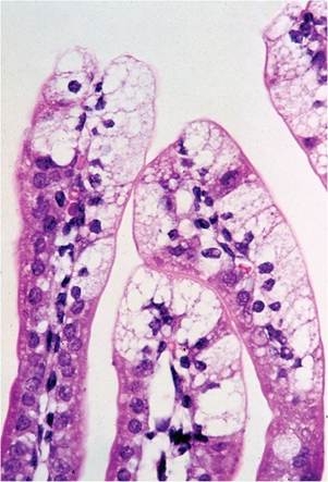
FIG. 1.43. Small intestine of an infant mouse infected with rotavirus. Note the cytoplasmic swelling of enterocytes at the tips of the villi. Microscopic lesions are often subtle and may not be readily apparent.
apical tubular system that occur in the distal small intestine, which may contain solitary eosinophilic globules. Definitive diagnosis can be achieved by electron microscopy of intestinal mucosa or feces. Rotavirus antigen can be detected in feces by ELISA, and RNA can be detected by PCR. Antigen detection can be accomplished with commercially available rotavirus diagnostic kits, but false-positive reactions can occur with certain mouse diets. Careful controls are therefore advised. Serology for EDIM virus antibody is useful for surveillance and retrospective confirmation of infection.
More on the topic Rotavirus Infection: Epizootic Diarrhea of Infant Mice:
- Rotavirus Infection: Infectious Diarrhea of Infant Rats
- Pneumonia Virus of Mice Infection
- Pneumonia Virus of Mice (PVM) Infection
- Wild, and probably pet mice, may serve as hosts to numerous helminth species, but laboratory mice have a limited repertoire of helminth parasitisms, most notably pinworms and tapeworms.
- Clostridium perfringens: Epizootic Rabbit Enteropathy
- ROTAVIRUS INFECTIONS
- PERSISTENT DIARRHEA
- Diarrhea
- MALABSORPTION SYNDROMES (CHRONIC DIARRHEA)
- Diarrhea Cryptosporidium