Nervous tissue
Clearly, the functional cell in the sense of impulse transmission is the neuron. Diagrammatic examples of

Fig.
4.68. Diagram of smooth muscle. The upper panel illustrates the arrangement of smooth muscle cells in a longitudinal view. The spindle-shaped cells with a single central nucleus make a tightly compacted layer of closely adhering cells. The lower panel illustrates the appearance of cellular profiles cut in cross section (see dashed line). Since some of the cells would be cut near the center, these profiles would be relatively large ovals with the nucleus in the center. Other profiles would be smaller oval shapes because of sections through the more narrowed tapered ends of the cells.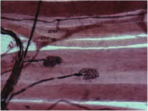
Fig. 4.70. Several motor endplates appear in this image. These motor neuron endings release acetylcholine to initiate depolarization and ultimately muscle contraction.
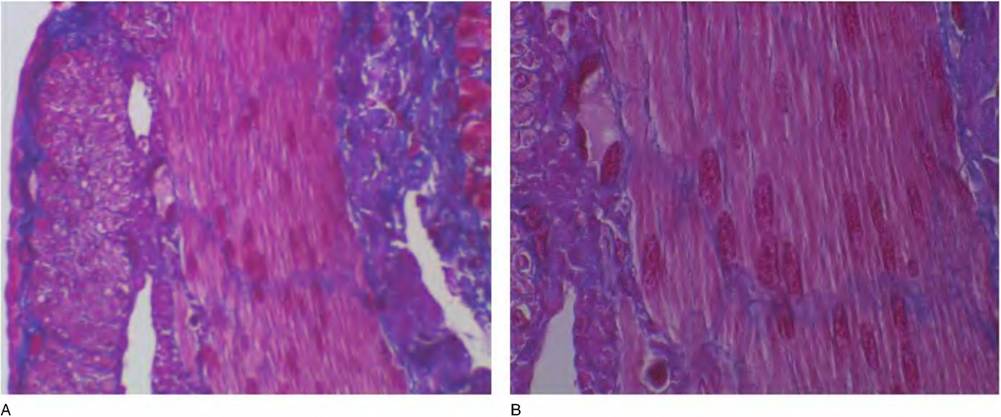
Fig. 4.69. Smooth muscle cells, small intestine. Panel A shows inner and outer layers of smooth muscle tissue in the muscularisexterna of the intestine. A small number of epithelial cells are visible to the extreme right of the image. The muscularisexterna (to the left of the figure) has an inner circular smooth muscle layer that has been sectioned longitudinally and an outer layer of cells that are oriented lengthwise along the intestinal tract. This cell layer has been cut in cross section. Panel B illustrates some of the cellular detail. Notice how the cells cut longitudinally seem to "flow" together. It is difficult to distinguish individual cells.
various types of neurons are illustrated in Chapter 8.
However, it is important to appreciate that neural tissue is more than simply collections of neurons. Figure 4.70 illustrates the interaction between the nervous system and the skeletal muscle. This image shows several motor nerve connections to skeletal muscle fibers. Figure 4.71 shows a cluster of myelinated nerve fibers that have been teased apart. The periodic pale areas that transverse individual nerve fibers are areas between Schwann cells (which create the myelin sheaths) called the nodes of Ranvier. These are regions where ion flow allows for rapid transmission (salutatory) impulse transmission in myelinated compared with nonmyelinated nerve fibers (see Chapter 8).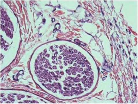
Fig. 4.72. This image illustrates portions of three nerve bundles (upper left, lower left, and lower center) cut in cross section. The analogy of nerve fibers like bundles of telephone cables passing through a conduit is apparent. Cross-sectioned blood vessels, adipocytes, collagen fibers, and fibroblasts appear in the surrounding area.
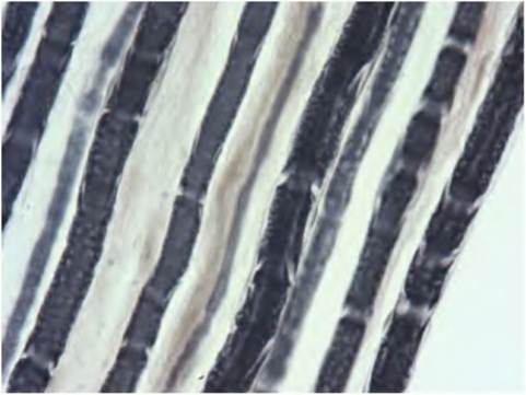
Fig. 4.71. These teased myelinated nerve fibers demonstrate the appearance of nodes of Ranvier (the pale areas that transverse individual fibers). These spaces between regions where Schwann cell growths to wrap the axons allow for saltatory nerve conduction, which is more rapid in myelinated compared with nonmyelinated nerve fibers.
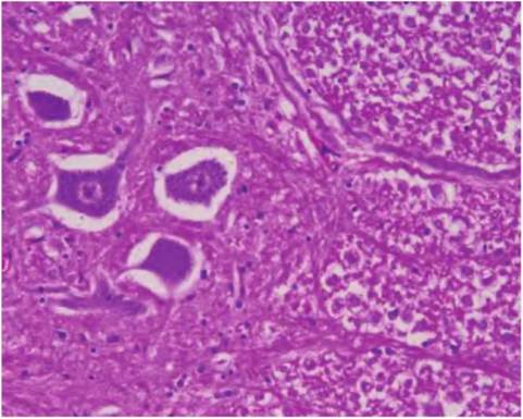
Fig. 4.73. Section of the spinal cord. A portion of the cell bodies of several motor neuron neurons appears to the left of the section. The dark spots are nuclei of smaller supporting cells.
Figure 4.72 shows a cross section through a nerve bundle. The analogy of nerves being like multiple phone lines bundled in a surrounding cable is apparent.
The health and functioning of neurons depends on many supportive cells called neuroglia or sometimes simply glia. Four types of neuroglia appear in the central nervous system and two in the peripheral nervous system. Some of these cells produce growth factors and other agents that promote health and tissue development, and others are responsible for- manufacturing the myelin sheath that acts to insulate many axons. The supporting cells of the CNS include astrocytes, microglia, ependymal cells, and oligodendrocytes. Astrocytes are important in anchoring neurons, and two of these cell types, Schwann cells in the peripheral nervous and oligodendrocytes in the central nervous system, produce the myelin sheath that wraps many axons (see Fig. 4.71). The structure of neurons related to the physics of conduction in nerves will be discussed in subsequent chapters. Figure 4.73 shows a spinal cord section to illustrate the nerve bodies of several large motor neurons. Because the axons do not necessarily appear on the same plane as the body of the nerve cell, it is rare to see a relatively thin two-dimensional tissue section that contains more a part of the cell body and perhaps a bit of the beginning (axon hillock)of the axon as it exits the cell body.
It is important to realize that this brief chapter is meant to simply give you some of the basic histology for each of the fundamental tissue types. We focused a great deal of effort on epithelial tissue because epithelial cells (of one organ or another) are responsible for the synthesis and secretion of nutrients, signaling molecules, enzymes, and so on; in other words, the functional attributes of many vital organs. However, it should be clear at this point that each of the basic tissues must interact for physiological function and homeostasis to be maintained. You should appreciate that the microscopic study of tissues and cells provides an important adjunct to better understand physiology. In short, as we indicated at the beginning, structure and function are ultimately intertwined at multiple levels. As you learn the attributes of various organs and organ systems, consider the organization, development, and differentiation of the cells within the organ that make the physiology possible.
More on the topic Nervous tissue:
- Nervous tissue
- Neuron structure
- LEVELS OF ORGANIZATION
- Muscle tissue
- Central nervous system cytology
- Classification of nerves
- Supportive cells
- Contents
- Cerebrospinal fluid analysis
- Microscopic Anatomy of Tissues