Rats are susceptible to a number of important bacterial pathogens. Recognizing that any scheme is imperfect and overlapping, the following text has been organized into 3 sections:
pathogens that are (i) primary enteric infections, (ii) primary respiratory infections, and (iii) other bacterial infections. Alphabetical listing is used within each category. This approach can best assist the pathologist with differential diagnoses.
Primary Respiratory Infections Bordetella bronchiseptica Infection
Bordetella bronchiseptica is an uncommon, typically opportunistic pathogen that has been associated with respiratory disease in laboratory rats. The organism is a more common inhabitant of the upper respiratory tract of species such as the guinea pig and domestic rabbit. The organism tends to colonize the apices of respiratory epithelial cells, resulting in impaired clearance by ciliated epithelial cells.
Pathology
Aerosol exposure to B. bronchiseptica in laboratory rats has resulted in lesions characterized by suppurative rhinitis, multifocal bronchopneumonia with polymorphonuclear cell and lymphocytic infiltration, and peribronchial lymphoid hyperplasia. In animals examined at 2 or more weeks postinoculation, there was fibroblast proliferation and mononuclear cell infiltration. Spontaneous cases of bronchopneumonia associated with B. bronchiseptica infection in rats feature a suppurative bronchopneumonia with consolidation of affected anteroventral areas of the lung. Frequently, there is an identifiable concurrent infection, such as rat coronavirus or Mycoplasma infection. Experimental infection of SPF rats resulted in transient infection without spread to contact rats, lending credence to coinfection as a critical element for natural disease.
Diagnosis
Isolation of the organism in large numbers from affected tissues is required for definitive diagnosis. Identification of copathogens should also be considered.
Cilia-Associated Respiratory (CAR) Bacillus Infection
Naturally occurring respiratory disease in rats has been associated with Cilia-associated respiratory (CAR) bacillus, a filamentous, argyrophilic bacterium with gliding motility that colonizes the ciliated epithelium of airways.
CAR bacillus has not been definitively classified, but it is closely related to members of the Flavobacter/Flexibacter group based upon 16S rRNA gene sequence analysis. The organism is Gram-negative and is difficult to grow on conventional cell-free media. It has also been demonstrated on respiratory epithelium in other species including mice, rabbits, cattle, goats, and pigs. Sequence analysis of 16S rRNA genes, antigenic comparisons, and experimental infectivity studies suggest that the rat CAR bacillus is closely related to isolates from other rodents, but distinct from the CAR bacillus of rabbits, cattle, and goats. CAR bacillus is associated with chronic respiratory disease (CRD) in the rat, frequently as a coinfection with M. pulmonis. Both CAR bacillus and M. pulmonis infect ciliated respiratory epithelium, resulting in perturbation of mucociliary clearance and development of CRD. Although CRD is often the result of coinfection with CAR bacillus and M. pulmonis, each of these agents can be found as single infections resulting in similar disease. CRD is often multifactorial, including environmental factors (ammonia) and viruses (Sendai virus). Although CAR bacillus was first described in 1980, there is evidence that the organism has been associated with respiratory disease in rats for decades, based upon retrospective staining of tissue sections. CAR bacillus is transmitted by direct contact, usually during the neonatal period. Experimental inoculation of various strains and stocks of rats (F344, LEW, and SD) has shown that rats are uniformly susceptible to CAR bacillus-induced disease, but CAR bacillus isolates differ in pathogenicity.Pathology
Infection may be subclinical with minimal or no microscopic lesions. Chronic suppurative bronchitis and bronchiolitis, with peribronchiolar cuffing with lymphocytes and plasma cells, are typical microscopic findings when disease is present. There is marked leukocytic infiltration in the lamina propria of affected airways.
Bronchiolec- tasis, with mucin and leukocyte accumulation, may result in bulging of the serosal surfaces of the lung, which is often asymmetrical in distribution (see “M. pulmonis infection”). Although organisms are discernable in H&E sections, they are particularly evident when stained with the Warthin-Starry method, revealing slender, argyrophilic bacilli inserted along the apices of the ciliated respiratory epithelium (Fig. 2.19).Diagnosis
The typical slender CAR bacteria are best demonstrated in tissue sections by silver stains, such as the Warthin-Starry stain. The organisms are also readily seen as electron- dense bacilli oriented between the cilia of respiratory epithelial cells by electron microscopy (Fig. 2.20). Serology can be used to detect seropositive animals. However, the use of CAR bacillus whole cell lysates as antigen for serology is problematic, in that this bacterium has a number of cross-reacting antigens among other bacteria. Other diagnostic techniques include the demonstration of the CAR bacillus in silver-stained tracheal scrapings using Steiner's silver stain, or in nasal swabs using PCR. The organism has been grown in embryonated chick eggs, cell culture, and, more recently, in cell-free media. Differential diagnoses include M. pulmonis infection and pneumonia due to conventional bacteria, with possible complications due to concurrent infections with other
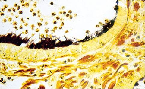
FIG. 2.19. Bronchiole from a rat infected with CAR bacillus. The cilia have been completely effaced by overgrowth of CAR bacilli (Warthin-Starry stain).
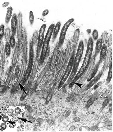
FIG. 2.20. Electron micrograph of bronchiolar epithelium colonized by CAR bacilli. Note the end-on attachment of the bacilli (arrowheads) among the cilia (arrows). The inset represents a cross section of bacilli among the cilia. (Source: Ganaway et al. 1985. Reproduced with permission from American Society for Microbiology.)
respiratory tract pathogens (Sendai virus, PVM, rat coronavirus, or Pneumocystis).
More on the topic Rats are susceptible to a number of important bacterial pathogens. Recognizing that any scheme is imperfect and overlapping, the following text has been organized into 3 sections::
- Rats are susceptible to a number of important bacterial pathogens. Recognizing that any scheme is imperfect and overlapping, the following text has been organized into 3 sections:
- APPENDIX 5 PATHOGENS SUSPECTED OF CAUSING WILD POPULATION DECLINES, OR OF CONSERVATION IMPORTANCE
- The foregoing sections feature many viral and bacterial infectious diseases of the gastrointestinal tract of rabbits, which often occur in combination and affect the overall enteric physiology of the rabbit in a syndrome known as dysbiosis.
- Wild rats are host to many nematodes that rarely infest laboratory rats, but there is ample evidence of wild rats serving as sources of laboratory rat infestations, generally through contamination of feed and bedding and occasionally through arthropod intermediate hosts, such as cockroaches.
- Neoplasia is very common in rats, and aged rats are often simultaneously afflicted with multiple types of neoplasia.
- 4 WILDLIFE PATHOGENS WITH ARTHROPOD VECTORS
- 3 SELECTED SOCIO-ECONOMICALLY IMPORTANT WILDLIFE RELATED PATHOGENS AND DISEASES IN EUROPE
- 2 SELECTED ZOONOTIC PATHOGENS WITH EUROPEAN WILDLIFE RESERVOIRS/HOSTS
- Epithelial Migration
- Article 4.11 Midsized issuers welcome funding scheme