EXAMPLES OF DISEASES CAUSED BY INSECTS, MITES, AND TICKS
Disease conditions are presented in the following section as somewhat isolated problems. However, health results from the combination of all factors influencing a host, and most parasites influence hosts in multiple ways.
Bloodfeeding ectoparasites can consume enough blood to cause anemia, and infestations can be associated with inflammatory dermatitis, alopecia (hair or feather loss) with resulting environmental exposure and ensuing energetic costs, annoyance, unthriftiness, chronic wasting, secondary bacterial infections, transmission of pathogenic agents, etc. Thus, the following conditions might best be thought of as participants in the cascades of interactions that influence health.Myiasis
Whereas some people are repulsed by thoughts of living animals parasitized by fly maggots, the natural histories of flies with parasitic larvae can be quite fascinating. For example, larvae of many species of flies feed on dead tissue and other organic debris found at the edges of wounds, and the feeding of such species may actually help to keep wounds clean by discouraging secondary bacterial infections. In fact, maggots of several species were used historically to treat human wounds before the advent of antibiotics, and the use of medical maggots is gaining in popularity again because antibiotic resistance of bacteria leaves physicians with few options for successful treatment (Sherman et al. 2000). Myiasis is the term used to describe such parasitism, and three general categories are used to describe infestations depending on the degree to which the fly species is dependent on host tissues: accidental myiasis, facultative myiasis, and obligatory myiasis (Scholl et al. 2009).
In accidental myiasis, also called pseudomyiasis, the maggots do not typically parasitize a living host; the host is just an accidental victim. Such accidental myiasis may occur when hosts consume vegetation or carrion infested with fly eggs.
These eggs may hatch and the surviving larvae may pass through the alimentary tract of the host. Larvae that persist in the individual host may cause considerable disease, but they are not typically associated with population-level problems for wildlife. Signs of disease associated with accidental infestations range from no apparent signs, through nausea, vomiting, and diarrhea, to more severe disease resulting from ulceration or perforation of the alimentary tract. In most cases, these larvae do not continue to develop or molt while in the host, so this is truly an accidental interaction (Scholl et al. 2009).In facultative myiasis, fly larvae feed either as saprophytes or as parasites, and they can be thought of as having the ability to opportunistically take advantage of many types of food sources including living flesh (Scholl et al. 2009). These species can be further characterized by their ability to initiate parasitism of living tissues. Primary facultative myiasis involves the invasion of living tissue through a small wound or through intact skin. Secondary facultative myiasis involves species that take advantage of wounds created by other maggots, but that can't invade a host without having access to a wound. Tertiary facultative myiasis involves those species of flies that invade a host at sites with primary and secondary species prior to host death, but that lack the ability to parasitize healthy hosts. Of course, nature is not limited to such neat categories, and some species of flies with maggots utilizing tertiary facultative myiasis will only rarely feed on living tissues, while others will do so commonly. Many species, including the blow flies, lay eggs on carrion in various states of decomposition. The evolutionary step is short indeed between a carrion feeder and one that feeds on hair matted with feces, urine, or blood or on dead tissues next to wounds. The next step, involving invasion of healthy tissues, is facilitated by the availability of healthy tissues next to such wounds or in association with soiled fur or feathers.
Thus, facultative myiasis can be thought of as an evolutionary transition between flies producing accidental myiasis and those whose larvae require living flesh to complete their life cycles (Scholl et al. 2009).North American species whose larvae commonly engage in some level of facultative myiasis include many species of the Families Calliphoridae [Chrysomya spp., Calliphora spp., Phormia regina, Cochliomyia macerllaria (the secondary screw worm), and Sarcophagidae (Sarcophaga spp.)]. Calliphorids, known collectively as blow flies or bottle flies, are moderately large flies with shiny metallic cuticles. Blow flies typically lay their eggs in carrion, but many species are attracted to purulent, necrotic wounds, chronic infections, or fur or feather soiled with urine or feces. Larvae feeding in such fetid conditions may invade healthy tissues as opportunity arises, and, if the host dies, overlapping generations of maggots may continue to feed on the dead body. Blow fly maggots drop away from the host prior to pupation in the leaf litter or loose dirt, and adults emerge approximately one week later to feed, breed, and continue their cycle (Scholl et al. 2009).
Fly larvae engaged in obligatory myiasis require the tissues of a living host for development to complete their life cycles. Such species include the primary screw worms, bot flies, warble flies, and “blood sucking maggots” (Protocalliphora spp.).
Cochliomyia hominivorax is known in North America as the primary screw worm (the Old World primary screw worm is Chrysomya bez- ziana). Females of this fly are attracted to wounds where they feed and lay eggs on the edges of such sites; screw worm flies commonly infest sites such as umbilical cords of newly born mammals, small wounds resulting from tick bites, and lacerations. Larval screw worms hatch in less than a day after oviposi- tion, and young larvae begin feeding in the exposed wound, enlarging it for themselves and for the next cohort of maggots.
The larvae develop quickly through their three instars, drop off of the host to pupate, and emerge as adults in as little as two weeks (Scholl et al. 2009). Such infestations tend to progress and can be highly invasive; death can result from screw worm maggots invading the body cavities and destroying internal organs, from secondary bacterial infections at the wound, or from subsequent septicemia. Moreover, this fly is an opportunistic generalist in that the species has been found on a large variety of wildlife, including Virginia opossums, cottontail rabbits, jackrabbits, American black bears, coyotes, white-tailed deer, pronghorn, and feral hogs, as well as pets, livestock, and people (Allan 2001a).The history of primary screw worms in the southern United States is an ecological story involving anthropogenic changes and an interesting method of eradication. By the early 1900s, cattle ranching and farming had altered the landscape of the southern United States, increasing the edge habitats available for deer and promoting the availability ofwoody legumes that are fed upon by the adult screw worm flies. The increased availability of altered habitats, the movement of cattle across long distances, and the increase in hosts available (including livestock) facilitated an increase in screw worm numbers and an extension of their geographic range (Strickland et al. 1981, Wobeser 1994). Attempts to eradicate the fly from the United States began during the mid-1950s. The strategy for eradication relied heavily on the fact that female screw worm flies mate once with a single male, and scientists reasoned that they could disrupt the fly's life cycle by blocking the successful fertilization of the next cohort of eggs. Disruption was accomplished by rearing screw worms on artificial substrates and irradiating them to produce sterile male flies. It was important that mass rearing and irradiation did not cause loss of male sexual activity and that females remained receptive to sterile males.
As many as 50 million sterile males were released from airplanes annually during the peak production of the program, achieving as high a density as 3,500 flies per km2 in Florida. Local populations declined quickly as the sterile males mated with wild females, and continued efforts eventually suppressed the species throughout the region. The initial goal was to release flies in the northern part of the geographic range and to continue releasing flies each year to push the northern border of the range to somewhere south of ranching interests in the United States. In areas where the species had been abundant, including most of Florida, Louisiana, Oklahoma, and Texas, losses to the livestock industry were estimated at over $100 million annually prior to eradication efforts (Allan 2001a). Although the species was not eradicated entirely, the suppression of screw worm populations allowed livestock ranching to thrive in the region. Not unexpectedly, such a program was very expensive, with a cost of $10 million annually in the 1950s (Strickland et al. 1981, Wobeser 1994, Allan 2001a). Although sterile male flies are still being used to suppress populations in Mexico, and to drive them further south through Central America, limited outbreaks do still occur north of the Mexican border (Wobeser 1994). Although the motivation for screw worm eradication had little to do with wildlife, many species, including white-tailed deer, benefited from the suppression of this deadly parasite.Screw worm flies were once considered the most important parasite of white-tailed deer in Texas and Florida, and, in some areas of Texas, fawn mortality was estimated to be as high as 80% during years of high fly densities, whereas only 25% died during years with low fly density (Strickland et al. 1981, Wobeser 1994, Allan 2001a). Clearly, the loss of 80% of fawns to a single mortality factor could limit populations of deer (Allan 2001a). Deer did not evolve with such significant losses to screw worms, because the increases in screw worm fly densities resulted directly from anthropogenic changes associated with cattle ranching.
Finally, suppression of screw worm populations in the southern United States was followed by a rapid expansion of deer populations (Strickland et al. 1981, Wobeser 1994), but continued increases in deer populations throughout the eastern United States suggests that many other factors (including changes in land use patterns) are responsible for the continued increase in numbers.Wohlfahrtia spp. (including W. vigil and W. opaca in the New World and W. magnifica in the Eurasia) are obligatory parasites in northern latitudes. Although rabbits and rodents are considered to be the primary reservoirs, infestations have been reported from a wide range of hosts, including cottontail rabbits, jackrabbits, rodents, mustelids, canids, domestic pets, waterfowl, and humans (Craine and Boonstra 1986, Allan 2001a). Eggs of these species develop and hatch within the females' bodies, and larvae are larviposited onto moist body openings, fresh wounds, or unbroken skin. The larvae then penetrate and create boillike cysts in the subdermis with a single opening in the skin through which many individual maggots breath. These species tend to parasitize young animals. Feeding by as few as four W. vigil maggots has been associated with mortality of animals as large as fox pups (Craine and Boonstra 1986, Allan 2001a), and this parasite can have significant impacts on host numbers. In one study, four of 43 nests of meadow voles, Microtus pennsylvanicus, were found to be infested with W. vigil (Craine and Boonstra 1986). Nestling voles were found moribund or dead infested with a mean of 10.8 (±1.5 SE) maggots per vole, with extensive pathology involving as much as 30% of hosts' tissues (Craine and Boonstra 1986). While the description sounds dramatic, such losses tend to be local in scope and typically do not limit vole populations.
Botflies and warble flies (Family Oestridae) represent another type of obligatory myiasis, and all species are parasites of mammals. There are many species of such flies, including the rodent and rabbit bots (Cuterebra spp.), warble flies of reindeer and caribou (Hypoderma tarandi), nasal bots of deer (Cephenemyia spp.), and the poetically named blood-sucking maggots (Protocal- liphora spp.). The life cycles of the flies presented are superficially similar, but differ in the biology of the species involved, the diseases they produce, and the implications for wildlife management.
Rodent and rabbit bots are maggots of Cuterebra spp. (including C. lepusculi and C. horripilum of rabbits, C. emasulator of a variety of rodents including tree squirrels, chipmunks, and deer mice in eastern North America, C. latifrons of woodrats, and C. fontinella and C. jellisoni of rodents in western North America). The adults are large, colorful, robust, bee-like flies with large eyes and vestigial mouthparts (adults do not feed); lack of functional mouthparts results in adults that live only long enough to mate, develop eggs, and oviposit. Thus, all of the nutrition necessary to complete the life cycle must be obtained during the period of the larval instars (Colwell 2001).
Breeding of Cuterebra spp. most commonly involves emergent males swarming over a prominent feature in the environment such as a hill top or large rock outcrop; females that fly through such swarms are quickly caught and mated. Females are univoltine (one generation per year) and oviparous; 1,000 to 4,000 eggs are oviposited on vegetation or substrate near the nests or warrens of their preferred hosts during summer months. After 5-7 days, the eggs hatch in response to higher temperatures and CO2 concentrations that often indicate presence of a host. First stage larvae crawl and attach to hair of the passing host, enter the body cavities via the eyes, nares, or through a fresh skin wound, and then migrate to the nasal passages, esophagus, or trachea. Second stage larvae continue to wander until they reach the preferred site, depending on the species, in the subdermal tissues (under the skin), where they cut a small hole in the host's skin. The larvae obtain nourishment by absorbing available host fluids while keeping their posterior ends, containing the respiratory spiracles, lodged against the breathing hole. The host reacts by surrounding the larva with a granulomatous wall or “warble” characteristic of bot fly infestations. Such lesions are most commonly observed in late summer or autumn months of August, September, and October. After growth through four successive molts, the mature larva squeezes through the breathing hole, drops to the ground, and burrows into the leaf litter or loose soil, where it pupates. Pupal diapause allows species in northern latitudes to survive until the combination of environmental characteristics and host availability is right for emergence of the adults (Cogley 1991, Colwell 2001).
The impact of cuterebrid maggots on their hosts was initially assumed to be great and has been somewhat controversial
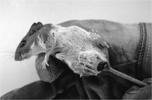
FIGURE 5.7 Cuterebra sp. bots in the groin and perineal region of a cotton mouse, Peromyscusgossypinus (Mullen and Catts 2002, copyright © Elsevier, by permission).
(Cogley 1991, Colwell 2001). In fact, C. emascu- lator, a parasite of mice and tree squirrels, was named because it was thought originally to feed on the testicles of its host. These larvae sometimes are found in subcutaneous tissues of the groin (Fig. 5.7), and their large size appears to displace the testicles, but they do not emasculate their hosts. Bot fly larvae obtain nutrients for growth and development from their hosts, and the energy depletion may impact survival or reproduction of individual hosts during times of resource scarcity. Although rare, larvae occasionally may migrate to abnormal tissues such as the nares or brain, where they may debilitate or kill individual hosts. Likewise, secondary bacterial infections may be associated with bot fly infestations, but other species of bots produce bacteriostatic secretions limiting abscess formation (Beesley 1968). Although it may seem counterintuitive, these maggots only rarely cause serious disease, and impacts on mean fitness or populations appear to be minimal.
Hypoderma tarandi is a fairly large fly with a dense covering of yellow and black setal hairs. Adults of these flies, as with other oestrids, lack functional mouthparts and therefore do not feed as adults. In contrast to Cuterebra spp., and other oestrids that swarm over high points in their environment, male Hypoderma spp. swarm over low-lying areas such as dry stream beds or along roadways (Anderson et al. 1994, Colwell 2001). Following mating, females are known to travel long distances to find their hosts, especially when such hosts are migratory species such as caribou, reindeer, or wildebeest; the maximum flight range of female H. tarandi was estimated to be as far as 600-900 km (Nilssen and Anderson 1995, Colwell 2001). Females are oviparous and, unlike Cuterebra spp., the female lands on the host to oviposit. Eggs are attached firmly to the base of hair shafts via a unique structure at one end of the eggs, and females apparently select individual hair shafts on which to oviposit; the result is that eggs are securely attached and are even somewhat resistant to grooming (Colwell 2001).
First stage larvae crawl to the skin surface and regurgitate enzymes that digest collagen, aiding their penetration through the skin. Larvae then migrate through the subcutaneous tissues to eventually reach a location along the dorsum of the host. As with Cuterebra spp., a hole is cut in the skin through which the maggot breathes. As they grow and develop, the larvae keep their anteriorly placed mouth deep in the granulomatous warble. Once matured, larvae exit through the breathing hole and fall to the ground to pupate during late spring or early summer. Emergent adults breed, and the cycle continues prior to the onset of autumn. Individual reindeer can have as many as 2,000 larvae (ranging in size to 2.5 cm in length) scattered under the skin along their backs. Heavy infections cause damage to the skin and underlying tissues, allergic reactions, nutritional imbalances, secondary infections, and immunosuppression (Karter et al. 1992); Hypoderma spp. from cattle have been shown to secrete a protease enzyme that breaks down host complement (C3), decreasing the host's potential immunologic response, but this enzyme has not been reported from H. tarandi (Colwell 2001). On warm summer days, large swarms of this fly cause reindeer to seek out patches of snow without flies, and this behavior may limit their ability to forage efficiently under such circumstances. Although it seems clear that Hypoderma spp. adversely impact milk yields in cattle (Reist et al. 2002), and one might assume energetic costs for hosts that reduce their feeding in attempts to avoid the parasites, weight gains of reindeer have been unaffected by control of warble flies (Oksanen and Nieminen 1998).
Nasal bots (sometimes called head bots) of deer (Cephenemyia spp.) are robust flies approximately the size of honey bees. Females are viviparous and larviposit (squirt) first stage larvae onto the muzzle or into the eyes of deer without landing; these larvae then crawl through the mucous secretions and membranes into the eye socket, nose, or mouth to find their way to the nasal passages. These yellow to brown maggots use their mouth hooks to attach to the tissues in the nasal passages and the tonsilar pouches of mule deer, black-tailed deer, white-tailed deer, elk, and caribou. Unlike the warble flies and rodent bots that assume subcutaneous locations, nasal bots are not encapsulated by a granuloma. Maggots feed on blood and secretions in nasal passages, where they continue to grow, develop, and molt. When fully mature, the larvae migrate to the opening of the nares and drop to the ground to pupate during the following spring or early summer. Adults emerge from the pupae, mate, and continue the life cycle (Cogley and Anderson 1981).
The true impact of these maggots on deer is unknown. Fully mature bots may exceed 25 mm in length, and infested deer may harbor large numbers of such maggots. Although few clear signs of overt disease are associated with most infestations, annoyance, weight loss, and even death, associated with migration into the cranial cavity, have been reported (Colwell 2001). Surely, the maggots block the nasal passages, and their growth in confined spaces seems likely to produce annoyance and potentially pain.
Although only the maggots parasitize the hosts, it may actually be the indirect effects of the adults that cause population-level phenomena by causing avoidance behavior (Anderson 1975, Karter and Folstad 1989, Moerschel and Klein 1997, Anderson 2001). Deer may stand with their noses to the ground or buried into their sides, and (reportedly) try to run away from the buzz of the females' wing beats (Anderson 1975, 2001). Reindeer seek snow or ridge tops with cooler temperatures, stronger winds, and fewer flies. They spend more time walking and running and less time lying down and eating (Karter and Folstad 1989, Hagemoen and Reimers 2002). Reduction in the relative time spent grazing during short Arctic summers may affect body condition (and potentially overwinter survival and reproduction) of reindeer and caribou (Downes et al. 1986, Moerschel and Klein 1997). Moreover, the stress induced by oestrid flies and fly avoidance causes physiologic responses, including activation of opioid peptides that act as neurochemicals effecting a wide range of responses that influence immunologic function and fitness (Colwell 2001).
The final example of myiasis involves the larvae of Protocalliphora spp., which are aptly called blood-sucking maggots. Adults of the different species of Protocalliphora lay their eggs on nestling birds or in the nests near newly hatched nestlings. The fly eggs hatch within 24 to 48 hours. The larvae of most species crawl to a place in the nest near a nestling where they can feed intermittently on nestling blood or other fluids through the skin of the feet and legs. Other species have larvae that directly enter the nostrils or ears, and P. braueri larvae burrow through the skin of nestlings to continue to feed as internal parasites (Bennett 1957, Rogers et al. 1991, Whitworth and Bennett 1992).
Blood Loss and Hemolysis
Blood is an extremely important tissue that transports oxygen, nutrients, and biochemicals (including hormones) to other tissues, carries metabolic waste products away from tissues, and contains many of the cells and proteins important to the immune system. Blood loss can result in a number of problems for hosts, and anemia (lower than normal volume or percentage of red blood cells or hemoglobin) can occur acutely when the number of red cells drops precipitously or develops more slowly with chronic disease. There are many causes of anemia, including hemolysis (the internal rupture of red blood cells, as occurs with some toxins that distort red blood cells, making them susceptible to elimination by the spleen, or with infections by protozoa causing malaria, leucocytozoonosis, hemobartonellosis, etc.). Alternatively, anemia may develop from an inability to create red blood cells (as occurs with some types of cancers and iron deficiency) or more directly from loss of blood. Blood loss can be associated with many etiologies (causes) including trauma (wounds, car collisions, etc.), the rupture of aneurysms, and internal or external parasites that feed on blood. Although ectoparasites may seem too small in relation to the host to remove enough blood to cause problems, large numbers of blood-feeding ectoparasites can result in both debility and death under the right (or wrong) circumstances. We describe three examples: feeding by bloodsucking maggots, swallow bugs, and ticks.
The first example is of blood loss caused by blood-sucking maggots mentioned above under the heading “myiasis”. These are muscoid flies, related to house flies (Musca domestica), and various other species of Calliphorid blow flies (Calliphora spp., Phormia spp., Lucilia spp., etc.) that lay eggs, hatch as small larvae, undergo three larval instars as maggots, pupate, and then feed, mate, and oviposit another generation of eggs as adults. The genus is widespread in North America, and most nesting avian species, including songbirds, swallows and martins, woodpeckers, hawks, and owls appear to be susceptible to infestation by one or more species of Protocalliphora (Bennett 1957, Rogers et al. 1991, Whitworth and Bennett 1992). The effects of blood-sucking maggots on nestling survival range from unapparent effects to mortality and nest abandonment.
Mature larvae of Protocalliphora spp. are large parasites in relation to nestling size, varying from about 7 to 17 mm (1¼ to ¾ inch), and each larva can consume a considerable volume of blood. When nests are infested with hundreds of such larvae, the result can devastate the entire brood (Bennett 1957, Rogers et al. 1991, Whitworth and Bennett 1992). The most common effects on individual birds include those typical of blood loss: anemia, weakness, and reduced growth rates. However, maggots that invade the hosts also cause direct damage to tissues, and all species create wounds that promote secondary bacterial infections. Anemic, debilitated nestlings grow slowly, increasing the duration of time spent in the nest and the probability that nestlings will succumb to predation prior to fledging. In other instances, the presence of large numbers of blood-sucking maggots may cause the parents to desert the nest. One report noted that 5-10% of infested nestlings died of blood loss and others that survived to fledge may have been less resistant to diseases and more susceptible to predation than would be strong healthy fledglings (Rogers et al. 19 91). Mature larvae undergo metamorphosis as pupae in the environment. Adults that emerge near the beginning or middle of the nesting season may repeat the cycle. Others that emerge near the end of the songbird fledging period over-winter in the nests or under bark or cracks in downed woody debris and lay eggs on nestlings during the following spring.
Some species of sucking lice have been associated with blood loss. As an example, the African blue louse, Linognathus africanus, is non-native in North America and has been associated with anemia and death of Columbian black-tailed deer (Odocoileus hemionus columbia- nus) and white-tailed deer (O. virginina) fawns (Brunetti and Cribbs 1971, Foreyt et al. 1986, Durden 2001) in the western United States.
Swallow bugs, Oeciacus vicarious and Oecia- cus hirundinis, illustrate another example of blood loss. These insects are related to human bed bugs (Order Hemiptera, Family Cimici- dae), and, like bedbugs, they reside near places of rest and feed while the hosts are sleeping. Swallow bugs reside in cracks in the mud nests of cliff swallows [Petrochelidon (formally Hirundo) pyrrhonota], purple martins (Progne subis), and potentially other socially breeding species in the Family Hirundinidae. Like other hemipteran bugs, there are five nymphal stages and the adults, which develop through a life cycle that often matches the timing of their migratory hosts. Although bats and other birds sometimes use swallow nests in their absence, and probably provide occasional blood meals for the bugs, most populations of bugs in temperate zones are forced to survive the 7-9 months of host absence prior to the migratory return of their primary hosts, that is, swallows. Surviving bugs help to reestablish the bug colony during the early phases of nest occupancy, and numbers increase throughout the breeding season. Further, nymphal swallow bugs appear to be transferred between nests by birds. Eggs of Oeciacus spp. are glued to the outside of mud nests, and, in heavy infestations, large numbers of eggs plastered to the nests can be seen from a distance (Krinsky 2009a). Heavily infested nests may produce enough immature and adult swallow bugs to literally drain the nestlings of their life blood, causing reduced growth, early fledging, and sometimes death or abandonment, of entire broods (Chapman and George 1991, Loye and Zuk 1991). Of course, these are not the only blood-sucking nest parasites that take advantage of the colonial breeding swallows. There are also mites, mosquitoes, and several species of ticks, including Argas cooleyi (Acari: Argasidae), Ornithodoros concanensis (Acari: Argasidae), and Ixodes baergi (Acari: Ixodidae), are commonly associated with swallow nests.
Swallow bugs transmit at least one important virus, a togavirus (Alphaviridae) known as Buggy Creek virus, to the birds. Several authors have reported the prevalence of the virus among swallows (Hopla et al. 1993, Brown et al. 2001), but the impacts of the virus on populations of swallows is not well understood. Interestingly, both the bug population density and the prevalence of the virus in the birds are directly correlated with the swallow colony size (Brown et al. 2001). Such a correlation allows the prediction of ectoparasite populations and virus persistence at colonies of swallows, and it suggests that there are costs associated with colonial breeding that may be directly associated with colony size. When we think about the evolution of colonial breeding, the effects of parasites and disease transmission should be among the variables considered.
All ticks are parasitic, and they all suck blood. However, the winter tick, Dermacentor albipictus, is notorious for the ability of large numbers of ticks to cause severely debilitating anemia and even death of large ungulates. This tick parasitizes domestic livestock, deer, elk, bighorn sheep, and moose in many areas throughout North America, from northern Mexico to approximately 60° north latitude in Alaska and Canada, but the problem becomes most extreme for moose in southern Canada (Samuel et al. 1991, Allan 2001b, Samuel 2004).
Dermacentor albipictus is a ł-host tick; once larvae attach to a host they remain on that host for the remainder of their lives, only dropping off as fed females to oviposit more eggs into the environment. Especially in northern regions, this species follows a regular rhythm of seasonality with one generation per year in which adult females engorge between February and May, each producing thousands of eggs (up to 4,400!) that hatch into hungry larval “seed ticks” during autumn (Allan 2001b, Samuel 2004). Moose especially feed large numbers of
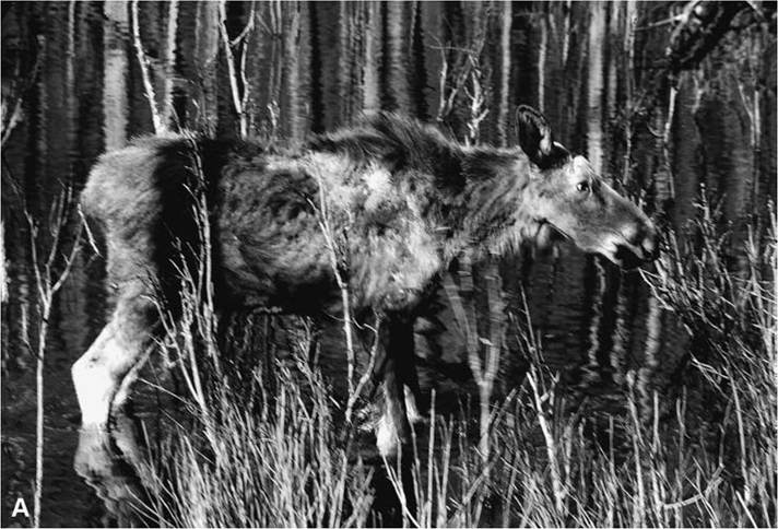
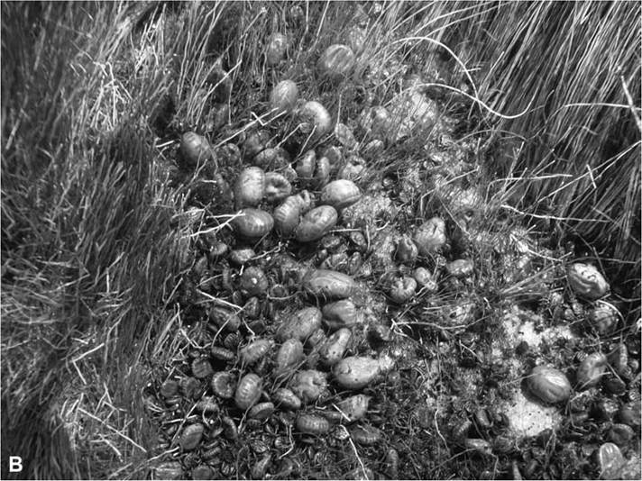
FIGURE 5.8 (A) A moose with alopecia and wasting associated with severe Dermacentor albipictus infestation and the condition known as ghost moose syndrome; (B) D. albipictus feeding on a moose with broken hair shafts (from Samuel 2004 and courtesy of Bill Samuel and Federation of Alberta Naturalists).
these ticks, and an “average” moose might harbor 33,000 to 50,000 ticks. Even higher numbers are found on some animals, including a bull with 150,000 ticks and a calf with 145,000 ticks (resulting in densities of 5.7 and 7.8 ticks per square centimeter or 37 and 50 ticks per square inch!) (Samuel 2004).
Summarizing decades of work on winter ticks on moose, Samuel (2004) estimated blood loss in moose as follows: bulls, cows, and calves with median numbers of ticks were found to feed 5,441, 3,161, and 8,123 female ticks per year, respectively. Each female tick was estimated conservatively to remove 1 gram of blood, equating to an estimated 5.4 L, 3.2 L, and 8.1 L of blood from the median bulls, cows, and calves, respectively. Such losses result in a required minimal replacement of 16.9%, 11%, and 57.9% of the blood volume of these median animals, respectively. Moreover, this blood loss occurs during the late winter and early spring, when temperatures are coldest, food is least plentiful, and thermoregulation afforded by a dense hair coat is most important (Glines and Samuel 1989).
Disease becomes apparent when heavy tick infestations cause annoyance and inflammation of the skin, resulting in grooming, scratching, and rubbing of the skin, and leading to damage of the hair coat and hair loss (alopecia). This combined nutritional and cold stress is exacerbated by the anemia caused by significant blood loss. Anemia causes animals to become weakened and lethargic, further reducing their ability to successfully forage and groom. Thus, weak animals become weaker, more ticks feed to repletion, hair loss and anemia worsen, sick animals become sicker, and some may die of malnutrition, exposure, decreased resistance to other parasites or diseases, or to reduced ability to avoid predation.
In Canada, severely affected moose are called ghost moose because damage and loss of the dark (brown or black) tips of the guard hairs exposes the inner hair shafts, which tend to be gray or whitish in color (Fig. 5.8). The appearance of such moose is often that of an undernourished animal with a ragged hair coat; the dorsal neck mane is often destroyed, there may be large patches of skin without hair or with only sparse and broken hair, and the shoulders and backs of the animals may have large gray to whitish patches (Samuel 2004). Although discussed here under blood loss, ghost moose syndrome would fit just as well with the next section.
Dermatitis
The skin is an amazing first defense against potential infectious agents, but it is not impervious. Cellular immune reactions to parasites and pathogens (helminths, protozoa, fungi, bacteria, and viruses) that colonize the skin surface, or in deeper layers, involve changes in the microvasculature and migration of white blood cells into the area. These changes result in the signs we all recognize as inflammation: tissues become swollen, red, hot, and painful (or itchy, depending on the severity); refer to Chapter 2. Some types of inflammation cause blisters to form, and sometimes ulceration of the epidermis exposes the deeper layers to secondary infections. Chronic inflammation can cause skin to become thickened (hypertrophied), dry, and flaky. Hair and feathers may become brittle and break, or they may fall out, get abraded from rubbing, or be plucked out. Hypersensitivity reactions to proteins in arthropod saliva often develop as a result of repeated bites of fleas, flies, bugs, lice, mites, or ticks. Such reactions may cause intense itching to the point of disrupting normal behavior and can even result in obsessive self-mutilation. Dermatitis varies from the inconsequential reaction to a single mosquito bite to the full-body loss of hair, with thickened, cracked, bleeding, infected, and unimaginably itchy skin associated with severe sarcoptic mange. Of course, the combination of chronic itching, hair or feather loss, and altered behavior are exacerbated by secondary bacterial infections at sites with cracked, ulcerated arthropod-bite wounds, or mutilated skin, as well as to exposure to environmental
conditions, chronic fatigue, blood loss, and transmission of parasitic organisms by biting arthropods. Examples of dermatitis induced by arthropods include hypersensitivity reactions to antigens associated with blood-feeding ectoparasites (such as sucking lice, kissing bugs, fleas, mosquitoes, tabanid flies, ticks, etc.), and the skin, feather, and hair damage associated with large numbers of chewing lice.
Two species of non-native chewing lice, Damalinia sp. and Bovicola tibialis, cause hair loss syndrome of deer in the western United States. Hair loss syndrome (H LS, or hair slip syndrome) caused by Damalinia sp. was first observed in populations of black-tailed deer in western Oregon and Washington in 1995 and had expanded south and east by 1998 (Bildfell et al. 2004). In 2005, another exotic louse, B. tibialis, was associated with HLS and mortality in central Washington, and both lice are now known to cause HLS as far south as the southern Sierra Nevada Mountains of California; and B. tibialis extends as far eastward as New Mexico and Texas (Westrom et al. 1976, Mertins et al. 2011). Hair loss syndrome presents as white or yellowish patches of fur of the thorax, neck, flanks, and rump of deer; discolored patches are coincident with the loss of guard hairs, revealing the under-fur. Hair loss may progress to loss of body condition, emaciation, and death. Hair loss syndrome typically causes more severe signs of disease in fawns than in adult deer, and loss of condition due to exposure has been associated with fawn mortalities that peak in late winter and early spring (Bender and Hall 2004, Bild- fell et al. 2004). The causes of death of fawns with HLS often seem associated with additional health problems, including lungworm pneumonia, nutritional deficiencies, and predation, and it may be that multiple causes commonly act synergistically to result in HLS deaths (Foreyt et al. 2004). Although prevalence of lice, and mortality from HLS fawns, may be high in some deer populations, at least one study has shown that over-winter survivorship of fawns in different management units did not correlate with prevalence of HLS (Bender and Hall 2004). Obviously, more work is needed to tease apart the relative importance of lice, HLS, and other mortality factors as influences of survivorship of deer fawns in areas with HLS (Krinsky 2009a). Most lice spend their entire lives on a single host, transmission of pathogens from host to host is rare, and relatively few disease agents important to wildlife populations are transmitted by lice (Durden 2004, Durden and Lloyd 2009). However, some lice do serve as important intermediate hosts of helminth parasites, including Trichodectes canis, an intermediate host of the double-pored tapeworm, Dipylidium caninum, of wild canids (Durden 2001, Durden and Lloyd 2009), and Trinoton anserinum, an intermediate host of the swan heart worm, Sarconema eurycerca (Seegar et al. 1976, Cohen et al. 1991).
Sarcoptic mange, or scabies, is caused by parasitism of mammals by the burrowing itch mite, S. scabei. The mite has been reported to cause disease in a large number of wild mammals, including marsupials, insectivores, rodents, lagomorphs, primates, carnivores, and ungulates (Bornstein et al. 2001, Pence and Ueckermann 2002, Mullen and O'Connor 2009). Mites in the genus Notoedres are superficially similar to S. scabei in morphology, disease produced, and general ecology, but they have a more narrow range of hosts; N. cati parasitizes mainly wild and domestic felids (Pence et al. 1982, 1995; Ryser-Degiorgis et al. 2002), and coatis (Valenzuela et al. 2000), and civets (Ninomiya et al. 2003), and N. cen- trifera causes disease in tree squirrels (Cornish et al. 2001). Development of clinical mange varies among species, by immune status of the host, and with population characteristics of the mites; it is intensely pruritic (causing intense itching) in most affected individuals, but some infested red foxes have failed to show signs of itching (Bornstein et al. 2001). Signs of disease typically include rash, dry, thickened, crusted, hyperpigmented skin, and alopecia. In addition, severe cases in wildlife may be associated with lymphadenopathy (swollen lymph nodes), inappetence, secondary bacterial infections, behaviors allowing approach by people, and death. Mangy wildlife typically appear unthrifty, with matted, odiferous hair coats, and thickened, crusty, hyperpigmented skin. In severe cases, alopecia (hair loss) may cover most or all of the body, and death likely results from exposure, secondary bacterial infections, and the energetic costs of fighting chronic disease. While epizootics of mange seem dramatic due to loss of life and the appearance of obviously diseased animals, Pence and Ueckermann (2002) point out that mange does not typically affect long-term population dynamics of self-sustaining populations of wildlife. Of course, small or threatened populations may suffer decimating losses when facing multiple insults, when loss of individuals affects population viability, when generalist parasites such as S. scabei are maintained by a community of hosts, and when researchers facilitate transmission through a diseased population (Cornish et al. 2001, Pence and Ueckermann 2002).
Psoroptes spp. are astigmatid mites that live in the ears of rabbits and ungulates, but they may spread out over the neck, shoulders, torso, and flanks when mite populations are unabated. Several species of Psoroptes were originally characterized by the host from which they were collected; Psoroptes cuniculi from rabbits, Pso- roptes cervinus from elk and deer, Psoroptes ovis from sheep, Psoroptes equi from horses, and so on. However, the validity of these species has been questioned and the need for taxonomic revision has been noted (Boyce and Brown 1991, Ramey et al. 2000, Zahler et al. 2000). A recent molecular comparison of Psoroptes spp. mites from different host species did not reveal species-specific patterns, and Zahler et al. (2000) suggested that all of these mites be considered conspecifics under the binomial associated with the earliest species description, that of P. equi. Even so, there remains enough population-level adaptation that mites may be unable to survive transfer to hosts of different species.
The life cycle of the astigmatid mite P. equi (including all related strains or subspecies) is interesting in that it depends, in part, on the host response, and the disease that results varies from minor annoyance to lifethreatening illness. Many generations of these mites occur on a single host animal, and the hypersensitivity response stimulated during feeding on host fluids results in serum being exuded through the skin. The mites live on the skin surface, feeding on the serum and dead cells. The serum exacerbates local inflammation, causing ulcerated lesions that result in scabs over the lesions. Scabs provide cover for the dense mat of mites, protecting them from the elements and the grooming of the host, as well as a providing a constant food supply (Mullen and O'Connor 2009). Transmission from host to host occurs with intimate contact (Mullen and O'Connor 2009). However, mites also survive off the host (in bedding, shed fur, etc., and depending on conditions) for as long as 10 to 15 days, facilitating potential indirect transmission (Meintjes et al. 2002). The extent of the scabby lesions, and associated hair loss, depends on the density of mites present, specific characteristics of the population of mites, and the hypersensitivity response of individual hosts. Some animals may harbor only a few scab mites in their ears with little apparent pathology. Others develop scabs in their ears that have been shown to block hearing, and loss of hearing might be expected to be associated with increased risks of predation (Norrix et al. 1995). Still other hosts succumb to exposure or secondary bacterial infections resulting from the combination of scab formation and hair loss over large areas of their bodies. Domestic sheep are often carriers of P. equi (subsp. ovis), and contact between domestic sheep and bighorn sheep has been responsible for die-offs of the latter in the western United States (Mullen and O'Connor 2009).
The alopecia and scabs resulting from heavy infestations of P. equi are sometimes referred to as scabies (as with “bighorn scabies”). In wild ungulates, severe disease has been reported among bighorn sheep (Lange et al. 1980, Welsh and Bunch 1983, Boyce and Brown 1991, Mazet et al. 1992, Singer et al. 1997), but infestations of mule deer, white-tailed deer, elk, ibex, and pronghorn also occur (Wright and Glaze 1988, Garris et al. 1991, Samuel et al. 1991, Ziccardi et al. 1996, Singer et al. 1997, Yeruham et al. 1999, Mullen and O'Connor 2009). In most infestations, mite populations are limited to the ears, resulting in few signs of overt disease. In mild cases in deer, inflammation may lead to mild bleeding and oozing of serum; such mild disease may be limited to small, discrete crusts on the inner surfaces of the ear pinnae and auditory canal. Indeed, where populations of white-tailed deer in the southeastern United States commonly suffer only mild disease, crusty lesions and inapparent infestations were observed with up to 80% prevalence (Roller et al. 1978, Garris et al. 1991). When cases progress to larger crusts with hair loss in and around the ears, the inflammation and pruritus may produce behavioral signs associated with head shaking, scratching, and rubbing on objects. In more severe cases, mite populations, and the inflammatory scabs they create, promote secondary bacterial infections. Ears may become filled with purulent discharge and infections may spread to the inner ear. Inner ear infections cause obvious changes in behavior, circling, loss of balance, and incoordination, and may result in death. Otitis externa (infection of the outer ear canal by bacteria and fungi) caused by mite-induced inflammation has been associated with occlusion of the ear canal of bighorn sheep (Norrix et al. 1995), and the subsequent reduction of hearing led the authors to logically suggest a connection between loss of hearing and increased susceptibility to predation. More commonly, as populations of mites grow large, they spread out from the ears over the trunk and body. Chronic severe infestations may lead to environmental exposure, weight loss, anemia, secondary infections, and death (Mullen and O'Connor 2009), but the chronic carrier state is common in most infested populations of wild ungulates.
Mites in the Family Sarcoptidae are more invasive than both P. equi and the fur mites previously discussed. These species also live for generations on individual hosts, but they are not limited to the surface of the skin. Sarcoptes scabei and Notoedres spp. are burrowers that excavate tunnels under the skin of mammals. Sarcoptes scabei infests a wide variety of wild mammals, including carnivores, ungulates, primates, and bats, and has worldwide distribution. Many researchers have commented on the potential number of species of the genus Sarcoptes associated with different host species, but others have advocated for one highly variable species (Bornstein et al. 2001). A molecular comparison of 23 isolates of S. sca- bei collected from four continents from dogs, foxes, lynx, raccoon, camel, chamois, a wombat, and livestock supported the existence of a single, variable species (Zahler et al. 1999). However, mite populations adapt to host species, and most transmission, which occurs via direct contact, is thought to be intraspecific rather than between species (Mullen and O'Connor 2009).
Male and female S. scabei breed on the skin of the host, and fertilized females burrow into the epidermis, depositing eggs in the burrows. Females lay up to three eggs per day for a period of 2-3 weeks before dying (Mullen and O'Connor 2009). Eggs hatch into larvae after 3 to 8 days, and larvae migrate to the surface to molt twice before maturing into adults. The entire life cycle requires only 14 to 21 days (Mullen and O'Connor 2009). Although a large majority of mites live on the surface, females defecate and die in the burrows, and the egg capsules remain in the burrows after the larvae hatch. Thus, considerable antigen remains in the burrows to initiate a hypersensitivity response that creates intense itching, and this hypersensitivity response to chronically present parasite antigen can be overwhelming for individual animals. It is the host's response, and not the mites per se, that causes the pathology associated with sarcoptic mange (Mullen and O'Connor 2009). Disease caused by
Notoedres spp. is similar to mange caused by S. scabei, but these mites are associated with felids, civets, squirrels, porcupines, and a number of Old World species of rats and mice. Mange was discussed further under the section on dermatitis.
The Families Knemidocoptidae and Epi- dermoptidae include several genera of mites superficially similar to S. scabei and Notoedres spp., but mites in these families cause mange in birds instead of mammals. Like S. scabei, these mites live on the skin, under scales, or in feather follicles. Fertilized females burrow through the epidermis, lay eggs in the burrows, and newly hatched larvae return to continue the cycle on the surface of the skin. Hypersensitivity reactions lead to thickened, crusty skin; scaly-leg; scaly-face; and small or large areas of feather loss.
Finally, the Family Oribatidae includes free- living mites that feed on organic detritus. These mites do not directly cause disease in wildlife, but some species serve as intermediate hosts of tapeworms, including the anoplocephalid Moniezia expansa. Definitive hosts accidentally ingest these small mites along with vegetation. Later, after the tapeworms have matured and eggs have been shed with host feces, oribatid mites feeding at the soil surface may ingest tapeworm eggs as they feed on organic debris (Mullen and O'Connor 2009).
Annoyance
In addition to the direct disease discussed above, ectoparasites likely cause considerable irritation and annoyance ofwildlife. “Fly worry” has been a known cause of decreased weight gains and milk production in cattle (Stork 1979, Jordaan and van Ark 1990, Wieman et al. 1992, Jonsson and Mayer 1999, Campbell et al. 2001, Cilek and Hallmon 2005). Such decreases in weight gain or milk yield occur because cattle feed less and expend energy avoiding flies when large densities are present. Such arguments have been extended to reindeer that clearly avoid swarms of biting flies during Arctic summers (Karter and Folstad 1989, Nilssen and Anderson 1995, Hagemoen and Reimers 2002). Reindeer and caribou seek snowdrifts, windy ridges, water, or other areas when fly densities are high. When unable to seek habitats with fewer flies, they may run to avoid the biting swarms. However, decreased weight gain of reindeer is not as clearly associated with annoyance as is that of cattle (Oksanen et al. 1993). Columbian black-tailed deer have been shown to behaviorally avoid flying female Cephenemyia spp. (Anderson 1975, 2001). However, the energetic impacts of avoidance of flies on the fitness of deer are not clear. While it seems logical that dense populations of ectoparasites could cause enough annoyance or irritation to reduce fitness of heavily infested individuals, such effects may be subtle, and the demonstration remains elusive. Annoyance may be expected to impact individuals during any given season, and to affect population dynamics for short periods of time under severe circumstances, but annoyance should not be thought of as a limit to populations of wildlife.
Toxicosis
Toxicosis results from the envenomization of wildlife by spiders, scorpions, ants, bees, and wasps; the poisonings related to consumption of poisonous insects, and toxins in the saliva of some ectoparasites. In general, toxicosis may cause disease in individual wildlife but does not impact populations. Two of the more interesting types of toxicosis result from attachment of specific species of ticks.
Tick paralysis results from neurotoxic proteins in the saliva of specific species of Ixodes, Dermacentor, Amblyomma, Rhipicephalus, Argas, and Ornithodoros ticks. In fact, at least 46 species of ticks in 10 genera have been associated with tick paralysis (Nicholson et al. 2009). In the United States, paralysis is caused by D. andersoni in the northwestern and Rocky Mountain areas, and D. variabilis in the eastern regions. In Africa, R. evertsi and O. savignyi cause paralysis in livestock and wildlife. Paralysis most commonly affects dogs, cats, and poultry. However, other species, including livestock, wildlife, and people, also are reported to suffer occasionally from tick paralysis; such events in wildlife are likely to be grossly under-reported.
Paralysis has been best described in relation to the bite of I. holocyclus in Australia, from which a salivary toxin has been isolated and described (Stone et al. 1989), and the protein causing paralysis appears to be somewhat similar among different species of ticks (Crause et al. 1994). Most cases result from large female ticks feeding on small or young animals. The signs of disease typically begin as weakness and incoordination of the limbs. As the toxin continues to affect the host, ascending flaccid paralysis progresses eventually to cause failure of the heart or diaphragm, followed rapidly by death. Removal of the tick from animals suffering tick paralysis may result in rapid improvement in people, pets, livestock, and wildlife, but cases caused by I. holocyclus may take weeks or months to resolve (Nicholson et al. 2009). Tick paralysis in wildlife has been reported in a western harvest mouse (Botzler et al. 1980), a red fox (Little et al. 1998), and at least 64 species of songbirds (Luttrell 1997). Wildlife succumbing to tick paralysis may be difficult to detect and may often die from other causes, such as predation; thus, cases of tick paralysis in wildlife are rarely reported.
Tick toxicosis is a similar response to unrelated proteins in tick salivary gland secretions. The disease manifests as an acute, febrile illness ranging in scope from localized inflammatory reactions, as is often associated with the bite of I. pacificus in the western United States and of I. holocyclus in Australia, through moderately severe, potentially life-threatening reactions to bites of the soft tick O. savignyi in Africa (Mans et al. 2003). Life-threatening systemic reactions, called sweating sickness of livestock and wild ungulates, are associated with the bites of Hyalomma spp., especially
H. truncatum females, in Africa. In the most severe cases, sweating sickness presents as sudden fever, followed by a wet rash over the body, necrosis of the oral mucosa, sloughing of hair and skin resulting in painful, ulcerated wounds prone to secondary bacterial infections causing emaciation, dehydration, depression, and death. Animals that survive are protected from future toxicosis by an immune response to the toxin.
Arthropods as Vectors
Concepts of population persistence, metapopulations, and the importance of dispersal routes connecting semi-isolated or small subpopulations are familiar to most students of wildlife biology and management. With our increasingly patchy, fragmented landscapes, humans intensify the need for corridors to promote gene flow and to provide a rescue effect for populations of wildlife that are slowly going extinct. Parasites also face such obstacles to individual survival and population persistence, and one of the most crucial of obstacles to parasite population persistence is that involving dispersal between hosts.
The vector-borne diseases of humans and domestic livestock, including the zoonotic diseases maintained in wild or domestic animal reservoirs, are explained in a large, and ever-growing, scientific literature, and related topics are reviewed in general texts on zoonotic diseases and medical or veterinary entomology (Acha and Szyfres 2001, 2003a, 2003b; Marquart et al. 2004; Mullen and Durden 2009). Likewise, arthropod vectors transmit many agents causing wildlife diseases. Disease ecologists typically use the term vector in relation to arthropods, and the pathogens transmitted by arthropod vectors are referred to as being vector-borne. However, the term vector also can be applied broadly to any animal or even a fomite that transmits an infectious agent. Thus, it is generally helpful to define such terms when used, and we will restrict our discussion of vector-borne pathogens to those transmitted by arthropods.
The nature of the relationships of a pathogen to both the vector and the host determines the details of biological cycles necessary for transmission, and knowledge of such details is required for an understanding of, and potential management of, the cycles. Vector-borne pathogens can be transmitted vertically from a female vector to her offspring or horizontally among hosts or between hosts and vectors (Reisen 2009). Transovarially transmitted pathogens are transmitted from female arthropods to their eggs, with subsequent larvae amplifying the prevalence of the pathogen through the vector population, but this requires that the pathogen not kill the developing eggs or larvae. Pathogens that are transmitted within an individual vector from stage to stage (e.g., viruses or bacteria that persist in a larval tick through the molt to the nymphal stage) are referred to as being transstadially transmitted.
Phoretic transfer describes the situation where arthropods are moved between hosts by other arthropods; examples include mites and lice that hitchhike between hosts on ectopara- sitic flies. The term mechanical transmission describes the movement of parasites or pathogens between hosts by arthropods; in this case, the vector functions merely as transportation for the pathogen. In contrast, biological transmission describes situations in which parasites or pathogens undergo an obligatory increases in number or development within the vector; in biological transmission, the vector serves as a required host as well as an agent for transportation (Reisen 2009). Examples of mechanical vectors include cockroaches or flies that carry fecal bacteria on their legs or mouthparts to food that is then ingested along with the agents that cause giardiasis (Giardia spp.) and avian salmonellosis (Salmonella spp.). Likewise, the deer flies and horse flies that serve as vectors of Francisella tularensis (the cause of tularemia), and the mosquitoes that transmit the viruses that causes avian pox, are mechanical vectors of the pathogens listed.
There are many examples of parasites that serve as biological vectors. The vector-parasite interactions involving biological vectors and their parasites are necessarily more complex than the interactions between parasites and mechanical vectors. Examples of parasites transmitted biologically include the arboviruses (including West Nile virus and the western and eastern equine encephalitis viruses), Borrelia burgdorferi sensu lato (the group of related spirochetes that cause Lyme borreliosis), Yersinia pestis (the causative agent of plague), various rickettsial agents (including Anaplasma spp. that cause anaplasmosis in humans and other mammals), Plasmodium and Leucocytozoon spp. (which cause avian hemosporidiosis or avian malaria), Elaeophora schneideri (an arterial worm of ungulates), and heart worms in the genera Dirofilaria, Sarconema, and Cordolobia.
In biological transmission, the parasites must undergo development, multiplication, or both during the period between exposure of a biological vector to a parasite and the time when it becomes infective to a naive host; such requirements relate directly to the life history characteristics of the parasites. When only multiplication is required, the process is termed propagative transmission. When developmental changes are required, the process is termed cyclodevelopmental, or just developmental, transmission, and when both multiplication and development are required the process is referred to as cyclopropagative transmission (Reisen 2009). Thus, the arboviruses, B. burgdorferi, Y. pestis, and Anaplasma spp. all must multiply in their vectors prior to effective propagative transmission; filarial worms must develop in their vectors prior to reaching the infective L3 stage prior to successful developmental transmission; and the protozoa causing avian malaria must both develop and multiply in their vectors prior to transmission of infective sporozoites via cyclodevelop- mental transmission.
Biological transmission often requires that a vector remain infected and pass the infection on through either transstadial (stage to stage) or transovarial (from a female to her offspring) transmission. There are many different potential blocks to such transmission. For instance, insects have a membrane lining their gut, the peritrophic membrane, that effectively blocks parasitic infections of many potential insect vectors (Harwood and James 1979, Gullan and Cranston 2005, Mullen and Durden 2009). In those cases of salivary transmission of pathogens (such as occurs with West Nile virus, various heart worms mentioned above, and parasites that cause avian malaria), the parasites have adapted some mechanism to cross this membrane in order to move from the gut to the salivary glands. Agents transmitted by ticks, such as Borrelia spp., have evolved characteristics favoring their retention during the complete reorganization inherent in the transstadial molts (Allan 2001b, Nicholson et al. 2009).
Indeed, pathogens have evolved many creative ways to ensure their transmission among hosts. However, morphological features and life history characteristics of potential vectors also influence transmission in predictable ways (Black and Kondratieff 2004). Vectors that feed on the blood of multiple hosts during their life cycles, that feed on a wide range of host species, and that have greater dispersal abilities tend to serve as vectors for a greater diversity of pathogens than do those that feed on single hosts, that specialize on a single species (or related group of species), that feed on skin, feathers, hair, or dead tissue rather than blood, and those that have poor dispersal ability.
The mouthparts of vectors directly affect the intimacy of their association with host tissues (e.g., blood, skin, mucous membranes, or contamination of host foods). Insect mouthparts have evolved from a primitive (but complicated) apparatus involving oral and pre-oral structures that facilitate manipulation of food items, ripping and tearing of organic matter, sometimes mastication, and movement of food into the pharynx (Black and Kondratieff 2004). Chewing mouthparts of the cockroaches and the grasshoppers provide well-recognized examples; anatomical structures include the labrum (a flap-like, dorsal, sensory lip), the mandibles (paired, jaw-like structures that lie beneath the labrum), maxillae (paired structures used to manipulate and chew food items before they are moved into the pharynx), and the hypopharynx (a tongue-like lobe that helps to mix salivary secretions with the chewed food bolus and then to suck the bolus into the pharynx). These basic structures have been modified in a myriad of ways, but we can group the modifications into at least eight additional categories, including: (1) a reduction of mouthparts to a vestigial state in adult bot flies (Order Diptera; Families Oestridae, Cuterebridae, Gastrophili- dae, and Hypodermatidae); (2) chewing mouthparts of chewing lice; (3) piercing mouthparts of sucking lice (Order Phthiraptera; Suborder Anoplura); piercing mouthparts of true bugs (Order Hemiptera); (4) piercing mouthparts of fleas (Order Siphonaptera); (5) sponging mouthparts of some flies (Order Diptera);
(6) piercing/sucking mouthparts of some flies;
(7) slashing mouthparts of horse flies and deer flies (Order Diptera; Family Tabanidae); and
(8) the piercing, sucking mouthparts of butterflies and moths. In addition, tissue-feeding maggots (fly larvae) have a pair of external, anterior mouth hooks used as anchors and to scratch and tear tissue. Tissue removed by the mouth hooks is mixed with salivary secretions and pumped through the gut of the maggot (Black and Kondratieff 2004, Gullan and Cranston 2005).
Each type of mouthparts allows a parasitic insect to obtain nourishment from hosts, but the different types of mouthparts, and ancillary tissues including salivary glands, result in different host-parasite interactions. Thus, the chewing mouthparts of amblycerans and ischnocerans allow the lice to feed on dead skin, feathers, and hair, but in most cases they do not allow direct access to the blood vascular system. As expected, chewing lice don't transmit many blood-borne pathogens (Clayton et al. 2008, Durden and Lloyd 2009). The slashing mouthparts of deer flies and horse flies create painful bites and feeding episodes are often interrupted (Allan 2001a, Hall and Gerhardt 2009). As expected, these flies are very good at mechanically transmitting pathogens that don't require long periods of feeding for transmission, such as F. tularensis, the causative agent of tularemia. Tabanid flies also serve as good biological vectors for parasites such as the arterial worm Elaeophora schneideri.
The dispersal ability of different species of arthropods is determined largely by (1) the presence or absence of wings, (2) the ability to survive prolonged periods of time in the environment, and (3) the ability to use widely ranging hosts for long-distance dispersal (Harwood and James 1979). Many groups of parasitic arthropods, including lice, fleas, mites, and ticks, lack the ability to fly. These parasites typically move long distances on their hosts to colonize new populations. While these species may be good vectors, their dispersal ability is hampered by their lack of flight. Of course, many groups of insects have evolved wings. Within the Diptera, the tiny size and delicate wing structure of midges and gnats make long-distance dispersal unlikely. Other groups, including horse flies, blow flies, flesh flies, and tsetses, are very strong fliers and have correspondingly greater potential to disperse long distances. Strong-flying vectors have the ability to move long distances and move pathogens quickly among different populations. Some arthropods, such as the astigmatid mites, survive only for a relatively short time off their hosts, while others such as ticks and fleas survive for extended periods of time in the environment between meals.
Non-volant species of vectors that remain on hosts during prolonged bouts of feeding, or after feeding, have the potential to move pathogens over greater distances than those that restrict their feeding to short periods in areas restricted to nests or burrows. Nest- and burrow-dwelling ectoparasites are known as nidicolous species; they typically require a narrow range of environmental conditions associated with the nests or burrows to which they are adapted, and they feed on a narrow range of hosts; nidicolous species tend to be specialists.
In contrast, ectoparasites with a more general palate are less likely to be associated with specific types of nests or burrows and are more likely to be exposed to a wide range of host species and their parasites (Durden and R. 2009, Nicholson et al. 2009). Clearly, the range of hosts to which parasites are adapted and associated varies between specialists and generalists, and affects their ability to transmit disease agents. As an example, the Ixodes ricinus group of ticks is infamous because it includes four generalist species (I. ricinus in Europe,
I. persulcatus in northern Asia, I. pacificus in the western United States, and I. scapularis in the eastern and southern United States) that transmit a large number of pathogens among a wide range of species, including humans. In contrast, nidicolous specialists within the
I. ricinus complex (including I. affinis, I. cookei, I. jellisoni, I. minor, I. muris, and I. spinipalpis) are known to transmit relatively few pathogens, and then among a relatively smaller number of host species (Nicholson et al. 2009).
Specialists and generalists vary in their relative abilities to focus transmission within a reservoir community and to amplify infections beyond the reservoir. In the most extreme examples, such as sucking lice and some species of fleas and ticks, highly specialized ectoparasites feed mainly on the reservoir host species, and they can be very efficient vectors, causing high prevalence of disease among hosts. Examples of transmission by specialists include that of Buggy Creek virus transmitted by swallow bugs, and the heart worm Sarco- nema eurycerca, transmitted among swans and geese via the chewing louse intermediate host, Trinoton anserinum. In the case of Buggy Creek virus, nidicolous bugs feed mainly on swallows and martins, serving to focus transmission and as an over-winter reservoir of the virus at colony nesting sites (Hopla et al. 1993). In the case of this heart worm, the species of louse (Suborder Amblycera) is a relative specialist on geese and swans, and, unlike many chewing lice, bites the host and feeds directly on blood. The blood-feeding habit allows intimate association between the birds' blood and the lice in which the immature stages (microfilariae through the infective L3) of the nematode develop, and the louse serves as an intermediate host in an enzootic transmission cycle (Seegar et al. 1976).
Generalist feeders may also dilute the force of transmission by feeding on non-reservoir hosts (LoGiudice 2003); the prevalence of infection among the population of vectors may be diminished or remain low when non-reservoir hosts account for a great enough proportion of the blood meals of the vector population. Extreme cases of host dilution effects converge on a phenomenon that has been termed zooprophylaxis, in which proteins in the blood of specific species of hosts actually kill parasites within blood-feeding vectors (Matuschka et al. 1993, Lane and Quistad 1998, Kelly and Thompson 2000, Saul 2003). On the other hand, generalist vectors serve as bridge vectors, carrying pathogens to hosts beyond enzootic maintenance cycles, to amplification or deadend hosts. Bridging is exemplified by four related species of Ixodes ricinus-group ticks that carry pathogens from enzootic cycles (in which they may also serve as maintenance vectors) to a wide range of host species including humans (Brown et al. 2005, Nicholson et al. 2009).
The number of times that a vector feeds on different hosts during its life cycle, and the timing of those feedings, affects vectorial capacity (or potential). Some species of flies do not feed as adults (including bot flies in the Family Oestridae), and the adults of these species are not known to transmit pathogens to hosts. In contrast, horse flies and deer flies are intermittent feeders and may fly between several successive hosts before obtaining a full blood meal (Allan 2001a, Hall and Gerhardt 2009). Such a feeding pattern coupled with the pain and blood flow associated with their bites, as discussed above, greatly facilitate their ability to serve as mechanical vectors. Likewise, the vector potential of different species of ticks is directly related to the number of hosts encountered during their life cycle. Ticks that feed on a single host [ł-host ticks such as Rhipicephalus (Boophilus) spp. and D. albipictus] transmit relatively few microorganisms, whereas the 3-host ticks tend to be more efficient vectors and transmit most of the tick-borne disease agents (Allan 2001b, Nicholson et al. 2009).
Thus, some groups of arthropods are notorious for transmitting disease agents, while other groups can be closely associated with infected hosts without efficiently transmitting pathogens. Vectorial capacity is a measure of an arthropod species' ability to serve as a vector between individuals in a population or community of hosts. Vectorial capacity depends on many aspects of the host-vector-parasite interactions, including the temporal and spatial overlap of hosts and vectors, biological associations of arthropods and pathogens, and the biological adaptations of these arthropods facilitating their own propagation. Specifically, vectorial capacity is dependent on the vector:host ratio, the probability that the vector will feed on a given host species, the typical number of days between blood meals, the proportion of vectors that become infected from feeding on infective hosts, the daily survivorship of the vectors, the probability that the exposed vector will become infective to subsequent naive hosts, and the number of days required for the vector to achieve such infectivity (Fine 1981, Black and Salman 2004).
The ecology of vector-borne diseases also varies somewhat from directly transmitted diseases. Like hosts and parasites, vectors have their own niche requirements, and the incidence of vector-borne disease in wildlife populations tends to depend upon temporal and spatial overlap of hosts and vectors (Marquart et al. 2004). In temperate regions, the activity pattern of vectors tends to be seasonal, and such seasonality results in seasonal occurrence of vector-borne disease that coincides with the activity of the primary, or amplification, vectors. Thus, the incidence of West Nile fever in birds in the United States increases with mosquito activity during the spring and subsides with the cessation of mosquito activity during autumn. Specific seasonal patterns are explained in more detail in chapters addressing bacteria, viruses, protozoa, and helminths. Clearly, a detailed understanding of the vectorhost relationships and transmission ecology requires considerable information on hosts, pathogens, and vectors. Moreover, the ecological interactions that determine the variables listed are likely to vary among different communities of hosts, vectors, and pathogens, as well as through time.
LITERATURE CITED
Acha, P. N., and B. Szyfres. 2001. Zoonoses and communicable diseases common to man and animals. Vol. I: Bacterioses and mycoses. 3rd ed. Report No. 580. Pan American Health Organization, Washington, DC.
Acha, P. N., and B. Szyfres. 2003a. Zoonoses and communicable diseases common to man and animals. Vol. 2: Chlamydioses, rickettsioses, and viroses. 3rd ed. Report No. 580. Pan American Health Organization, Washington, DC.
Acha, P. N., and B. Szyfres. 2003b. Zoonoses and communicable diseases common to man and animals. Vol. 3: Parasitoses. 3rd ed. PReport No. 580. Pan American Health Organization, Washington, DC.
Adler, P. H., and J. W. McCreadie. 2009. Black flies (Simuliidae). Pp. 189-206 in G. R. Mullen and
L. A. Durden (editors), Medical and veterinary entomology. 2nd ed. Academic Press/Elsevier, San Diego, CA.
Allan, S. A. 2001a. Biting flies (Class Insecta: Order Diptera). Pp. 18-45 in W. M. Samuel, M. J. Pybus, and A. A. Kocan (editors), Parasitic diseases of wild mammals. Iowa State University Press, Ames, IA.
Allan, S. A. 2001b. Ticks (Class Arachnida: Order Acarina). Pp. 72-106 in W. M. Samuel,
M. J. Pybus, and A. A. Kocan (editors), Parasitic diseases of wild mammals. Iowa State University Press, Ames, IA.
Anderson, J. R. 1975. The behavior of nose bot flies (Cephenemyia apicata and C. jellisoni) when attacking black-tailed deer (Odocoileus hemionus columbianus) and the resulting reactions of the deer. Canadian Journal of Zoology 53:977-992.
Anderson, J. R. 2001. Larviposition by nasopharyngeal bot fly parasites of Columbian black-tailed deer: A correction. Medical and Veterinary Entomology 15:438-442.
Anderson, J. R., A. C. Nilssen, and I. Folstad. 1994. Mating behavior and thermoregulation of the reindeer warble fly, Hypoderma tarandi L. (Diptera: Oestridae). Journal of Insect Behavior 7:679-706.
Beesley, W. N. 1968. Observations on the biology of the ox warble-fly (Hypoderma: Diptera, Oestridae). II: Bacteriostatic properties of larval extracts. Annals of Tropical Medicine and Parasitology 62:8-12.
Bender, L. C., and P. B. Hall. 2004. Winter fawn survival in black-tailed deer populations affected by hair loss syndrome. Journal of Wildlife Diseases 40:444-451.
Bennett, G. F. 1957. Studies on the genus Proto- calliphora (Diptera: Calliphoridae). Ph.D. dissertation, University of Toronto, Toronto, Ontario, Canada.
Bildfell, R. J., J. W. Mertins, J. A. Mortenson, and D. F. Cottam. 2004. Hair-loss syndrome in blacktailed deer of the Pacific Northwest. Journal of Wildlife Diseases 40:670-681.
Black, W. C., IV, and B. C. Kondratieff. 2004. Evolution of arthropod disease vectors. Pp. 9-23 in W. C. Marquart, B. C. Kondratieff, C. G. Moore,
J. E. Freier, H. H. Hagedorn, W. C. Black,
A. A. James, J. Hemingway, and S. Higgs (editors), The biology of disease vectors. Elsevier Academic Press, Burlington, MA.
Black, W. C., IV, and D. Salman. 2004. Molecular techniques for epidemiology and the evolution of arthropod-borne pathogens. Pp. 227-255 in W. C. Marquart, B. C. Kondratieff, C. G. Moore,
J. E. Freier, H. H. Hagedorn, W. C. Black, A. A. James, J. Hemingway, and S. Higgs (editors), The biology of disease vectors. Elsevier Academic Press, Burlington, MA.
Bornstein, S., T. Morner, and W. M. Samuel. 2001. Sarcoptes scabei and sarcoptic mange. Pp. 107-119 in W. M. Samuel, M. J. Pybus, and A. A. Kocan (editors), Parasitic diseases of wild mammals. Iowa State University Press, Ames, IA.
Botzler, R. G., J. Albrecht, and T. Schaefer. 1980. Tick paralysis in a western harvest mouse (Reithrodontomys megalotis). Journal of Wildlife Diseases 16:223-224.
Boyce, W. M., and R. N. Brown. 1991. Antigenic characterization of Psoroptes spp. (Acari: Pso- roptidae) mites from different hosts. Journal of Parasitology 77:675-679.
Brown, C. R., N. Komar, S. B. Quick, R. A. Sethi,
N. A. Panella, M. B. Brown, and M. Pfeffer. 2001. Arbovirus infection increases with group size. Proceedings of the Royal Society of London B: Biological Sciences 268:1833-1840.
Brown, R. N., R. S. Lane, and D. T. Dennis. 2005. Geographic distributions of tick-borne diseases and their vectors. Pp. 363-391 in J. Goodman, D. T. Dennis, and D. Sonenshine (editors), Tick- borne diseases of humans. ASM Press, Washington, DC.
Brunetti, O., and H. Cribbs. 1971. California deer deaths due to massive infestation by the louse (Linognathus africanus). California Fish and Game 57:162-166.
Campbell, J. B., S. R. Skoda, D. R. Berkebile, D. J. Boxler, G. D. Thomas, D. C. Adams, and R. Davis. 2001. Effects of stable flies (Diptera: Muscidae) on weight gains of grazing yearling cattle. Journal of Economic Entomology 94:780-783.
Capinera, J. L. 2010. Insects and wildlife: Arthropods and their relationships with wild vertebrate animals. Wiley-Blackwell, Hoboken, NJ.
Chapman, B. R., and J. E. George. 1991. The effects of ectoparasites on cliff swallow growth and survival. Pp. 69-92 in J. E. Loye and M. Zuk (editors), Bird-parasite interactions: Ecology, evolution, and behaviour. Oxford University Press, New York.
Chilton, G., M. J. Vonhof, B. V. Peterson, and
N. Wilson. 2000. Ectoparasitic insects of bats in British Columbia, Canada. Journal of Parasitology 86:191-192.
Cilek, J. E., and C. F. Hallmon. 2005. The effectiveness of the mosquito magnet trap for reducing biting midge (Diptera: Ceratopogonidae) populations in coastal residential backyards. Journal of the American Mosquito Control Association 21:218-221.
Clayton, D. H., R. J. Adams, and S. E. Bush. 2008. Phthiraptera, the chewing lice. Pp. 515-526 in C. T. Atkinson, N. J. Thomas, and D. B. Hunter (editors), Parasitic diseases of wild birds. Wiley-Blackwell, Ames, IA.
Clayton, D. H., and K. P. Johnson. 2003. Linking coevolutionary history to ecological process: Doves and lice. Evolution 57:2335-2341.
Cogley, T. P. 1991. Warble development by the rodent bot Cuterebra fontinella (Diptera: Cuterebridae) in the deer mouse. Veterinary Parasitology 38:275-288.
Cogley, T. P., and J. R. Anderson. 1981. Invasion of black-tailed deer by nose bot fly larvae (Diptera: Oestridae: Oestrinae). International Journal for Parasitology 11:281-286.
Cohen, S., M. T. Greenwood, and J. A. Fowler. 1991. The louse Trinoton anserinum (Amblycera: Phthiraptera), an intermediate host of Sarconema eurycerca (Filarioidea: Nematoda), a heartworm of swans. Med Vet Entomol 5:101-110.
Colwell, D. D. 2001. Bot flies and warble flies (Order Diptera: Family Oestridae). Pp. 46-71 in W. M. Samuel, M. J. Pybus, and A. A. Kocan (editors), Parasitic diseases of wild mammals. Iowa State University Press, Ames, IA.
Cornish, T. E., M. J. Linders, S. E. Little, and
W. M. Vander Haegen. 2001. Notoedric mange in western gray squirrels from Washington. Journal of Wildlife Diseases 37:630-633.
Craine, I. T., and R. Boonstra. 1986. Myiasis in Wohlfahrtia vigil in nestling Microtus pennsylvani- cus. Journal of Wildlife Diseases 22:587-589.
Crause, J. C., S. van Wyngaardt, R. Gothe, and A. W. Neitz. 1994. A shared epitope found in the major paralysis inducing tick species of Africa. Experimental and Applied Acarology 18:51-59.
Downes, C. M., J. B. Theberge, and S. M. Smith. 1986. The influence of insects on the distribution, microhabitat choice, and behavior of the Burwash caribou herd. Canadian Journal of Zoology 64:622-629.
Durden, L. 2004. Lice, the Phthiraptera. Pp. 67-76 in W. C. Marquart, B. C. Kondratieff, C. G. Moore, J. E. Freier, H. H. Hagedorn, W. C. Black, A. A. James, J. Hemingway, and S. Higgs (editors), The biology of disease vectors. Elsevier Academic Press, Burlington, MA.
Durden, L. A. 2001. Lice (Phthiraptera). Pp. 3-17 in W. M. Samuel, M. J. Pybus, and A. A. Kocan (editors), Parasitic diseases of wild mammals. Iowa State University Press, Ames, IA.
Durden, L. A., and N. C. Hinkle. 2009. Fleas (Siphonap- tera). Pp. 115-135 in G. R. Mullen and L. A. Durden (editors), Medical and veterinary entomology. 2nd ed. Academic Press/Elsevier, San Diego, CA.
Durden, L. A., and J. E. Lloyd. 2009. Lice (Phthirap- tera). Pp. 59-82 in G. R. Mullen and L. A. Durden (editors), Medical and veterinary entomology. 2nd ed. Academic Press/Elsevier, San Diego, CA.
Durden, L. A., and M. G. R. 2009. Introduction. Pp. 1-12 in G. R. Mullen and L. A. Durden (editors), Medical and veterinary entomology. 2nd ed. Academic Press/Elsevier, San Diego, CA.
Durden, L. A., and N. Wilson. 1991. Parasitic and phoretic arthropods of sylvatic and commensal white-footed mice (Peromyscus leucopus) in central Tennessee, with notes on Lyme disease. Journal of Parasitology 77:219-223.
Fahrenholz, H. 1913. Ectoparasiten und Abstam- mungslehre. Zoologische Anzeiger 41:371-374.
Fine, P. E. M. 1981. Epidemiological principles of vector mediated transmission. Pp. 77-91 in
J. J. McKelvey, B. F. Eldridge, and
K. Maramorosch (editors), Vectors of disease agents. Praeger Scientific, New York.
Foreyt, W. J., B. Hall, and L. Bender. 2004. Evaluation of ivermectin for treatment of hair loss syndrome in black-tailed deer. Journal of Wildlife Diseases 40:434-443.
Foreyt, W. J., D. H. Rice, and K. C. Kim. 1986. Pediculosis of mule deer and white-tailed deer fawns in captivity. Journal of the American Veterinary Medical Association 189:1172-1173.
Foster, W. A., and E. D. Walker. 2009. Mosquitoes (Culicidae). Pp. 207-259 in G. R. Mullen and
L. A. Durden (editors), Medical and veterinary entomology. 2nd ed. Academic Press/Elsevier, San Diego, CA.
Fritsche, T. R. 1999. Arthropods of medical importance. Pp. 1449-1466 in P. R. Murray, E. J. Baron,
M. A. Pfaller, F. C. Tenover, and R. H. Yolken (editors), Manual of clinical microbiology. American Society for Microbiology, Washington, DC.
Gage, K. L. 2004. Fleas, the Siphonaptera.
Pp. 77-92 in W. C. Marquart, B. C. Kondratieff,
C. G. Moore, J. E. Freier, H. H. Hagedorn, W. C. Black, A. A. James, J. Hemingway, and S. Higgs (editors), The biology of disease vectors. Elsevier Academic Press, Burlington, MA.
Garris, G. I., J. B. Prullage, J. L. Prullage, F. C. Wright, and J. A. Miller. 1991. Control of Psoroptes cuniculi in captive white-tailed deer with ivermectin-treated corn. Journal of Wildlife Diseases 27:254-257.
Glines, M. V., and W. M. Samuel. 1989. Effect of Dermacentor albipictus (Acaridxodidae) on blood composition, weight gain and hair coat of moose, Alces alces. Experimental and Applied Acarology 6:197-213.
Gullan, P. J., and P. S. Cranston. 2005. The insects: An outline of entomology. 3rd ed. Blackwell Publishing, Ltd., Malden, MA.
Hagemoen, R. I. M., and E. Reimers. 2002. Reindeer summer activity pattern in relation to weather and insect harassment. Journal of Animal Ecology 71:883-892.
Haldane, J. B. S. 1932. Possible worlds: And other essays. Chatto and Windus, London, UK.
Hall, R. D., and R. R. Gerhardt. 2009. Flies (Diptera). Pp. 137-152 in G. R. Mullen and L. A. Durden (editors), Medical and veterinary entomology. 2nd ed. Academic Press/Elsevier, San Diego, CA.
Halos, L., T. Jamal, Maillard, B. Girard, J. Guillot, B. Chomel, M. Vayssier-Taussat, and H.-J. Boulouis. 2004. Role of Hippoboscidae flies as potential vectors of Bartonella spp. infecting wild and domestic ruminants. Applied Environmental Microbiology 70:6302-6305.
Harwood, R. F., and M. T. James. 1979. Entomology in human and animal health. 7th ed. Macmillan Publishing Company, New York.
Hoberg, E. P., D. R. Brooks, and D. Siegel-Causey. 1997. Host-parasite co-speciation: History, principles, and prospects. Pp. 212-235 in D. H. Clayton, and J. Moore (editors), Hostparasite evolution: General principles and avian models. Oxford University Press, Oxford, UK.
Hoogstraal, H. 1985. Argasids and nuttallielid ticks as parasites and vectors. Advances in Parasitology 24:135-238.
Hopla, C. E., D. B. Francy, C. H. Calisher, and J. S. Lazuick. 1993. Relationship of cliff swallows, ectoparasites, and an Alphavirus in west-central Oklahoma. Journal of Medical Entomology 30:267-272.
Jongejan, F., and G. Uilenberg. 2004. The global importance of ticks. Parasitology 129(Suppl.):S3-14.
Jonsson, N. N., and D. G. Mayer. 1999. Estimation of the effects of buffalo fly (Haematobia irritans exigua) on the milk production of dairy cattle based on a meta-analysis of literature data. Medical and Veterinary Entomology 13:372-376.
Jordaan, L. C., and H. van Ark. 1990. A survey of annoyance of livestock by Simulium chutteri Lewis along the Orange River, South Africa (Diptera: Simuliidae). Onderstepoort Journal of Veterinary Research 57:189-195.
Jovani, R., J. L. Tella, D. Sol, and D. Ventura. 2001. Are hippoboscid flies a major mode of transmission of feather mites? Journal of Parasitology 87 1187-1189
Karter, A. J., and I. Folstad. 1989. Defence behaviour of reindeer in response to flying parasitic Diptera. Rangifer 9:14-16.
Karter, A. J., I. Folstad, and J. R. Anderson. 1992. Abiotic factors influencing embryonic development, egg hatching, and larval orientation in the reindeer warble fly, Hypoderma tarandi. Medical and Veterinary Entomology 6:355-362.
Kelly, D. W., and C. E. Thompson. 2000. Epidemiology and optimal foraging: Modelling the ideal free distribution of insect vectors. Parasitology 120:319-327.
Krinsky, W. L. 2009a. True bugs (Hemiptera).
Pp. 83-99 in G. R. Mullen and L. A. Durden (editors), Medical and veterinary entomology. 2nd ed. Academic Press/Elsevier, San Diego, CA.
Krinsky, W. L. 2009b. Tsetse flies (Glossinidae). Pp. 297-308 in G. R. Mullen and L. A. Durden (editors), Medical and veterinary entomology. 2nd ed. Academic Press/Elsevier, San Diego, CA.
Lane, R. S., and G. B. Quistad. 1998. Borreliacidal factor in the blood of the western fence lizard (Sceloporus occidentalis). Journal of Parasitology 84:29-34.
Lange, R. E., A. V. Sandoval, and W. P. Meleney.
1980. Psoroptic scabies in bignorn sheep (Ovis canadensis mexicana) in New Mexico. Journal of Wildlife Diseases 16:77-82.
Little, S. E., W. R. Davidson, E. W. Howerth, P. M. Rakich, and V. F. Nettles. 1998. Diseases diagnosed in red foxes from the southeastern United States. Journal of Wildlife Diseases 34:620-624.
Lloyd, J. E. 2009. Louse flies, keds, and related flies (Hippoboscoidea). Pp. 339-352 in G. R. Mullen and L. A. Durden (editors), Medical and veterinary entomology. 2nd ed. Academic Press/ Elsevier, San Diego, CA.
LoGiudice, K. 2003. Trophically transmitted parasites and the conservation of small populations: Raccoon roundworm and the imperiled Allegheny woodrat. Conservation Biology 17:258-266.
Loye, J. E., and M. Zuk (editors). 1991. Bird-parasite interactions. Oxford University Press, Oxford, UK.
Luttrell, M. P. 1997. Avian tick paralysis. In
W. R. Davidson, and V. F. Nettles (editors), Field manual of wildlife diseases in the southeastern United States. Southeastern Cooperative Wildlife Disease Study, College of Veterinary Medicine, The University of Georgia, Athens, GA.
Madslien, K., B. Ytrehus, T. Vikoren, J. Malmsten,
K. Isaksen, H. O. Hygen, and E. J. Solberg. 2011. Hair-loss epizootic in moose (Alces alces) associated with massive deer ked (Lipoptena cervi) infestation. Journal of Wildlife Diseases 47:893-906.
Mans, B. J., A. I. Louw, and A. W. Neitz. 2003. The major tick salivary gland proteins and toxins from the soft tick, Ornithodoros savignyi, are part of the tick Lipocalin family: Implications for the origins of tick toxicoses. Molecular Biology and Evolution 20:1158-1167.
Marquart, W. C., B. C. Kondratieff, C. G. Moore, J. E. Freier, H. H. Hagedorn, W. C. Black, A. A. James, J. Hemingway, and S. Higgs (editors). 2004. The biology of disease vectors. Elsevier Academic Press, Burlington, MA.
Matuschka, F. R., M. Heiler, H. Eiffert, P. Fischer, H. Lotter, and A. Spielman. 1993. Diversionary role of hoofed game in the transmission of Lyme disease spirochetes. American Journal of Tropical Medicine and Hygiene 48:693-699.
Mazet, J. A., W. M. Boyce, J. Mellies, I. A. Gardner, R. K. Clark, and D. A. Jessup. 1992. Exposure to Psoroptes sp. mites is common among bighorn sheep (Ovis canadensis) populations in California. Journal of Wildlife Diseases 28:542-547.
Meintjes, T., L. J. Fourie, and I. G. Horak. 2002. On- host ecology and off-host survival of the sheep scab mite Psoroptes ovis. Onderstepoort Journal of Veterinary Research 69:273-283.
Mertins, J. W., J. A. Mortenson, J. A. Bernatowicz, and P. B. Hall. 2011. Bovicola tibialis (Phthirap- tera: Trichodectidae): Occurrence of an exotic chewing louse on cervids in North America. Journal of Medical Entomology 48:1-12.
Moerschel, F. M., and D. R. Klein. 1997. Effects of weather and parasitic insects on behavior and group dynamics of caribou of the Delta Herd, Alaska. Canadian Journal of Zoology 75:1659-1670.
Moon, R. D. 2009. Muscid flies (Muscidae).
Pp. 275-295 in G. R. Mullen and L. A. Durden (editors), Medical and veterinary entomology. 2nd ed. Academic Press/Elsevier, San Diego, CA.
Morse, S. F., K. J. Olival, M. Kosoy, S. Billeter,
B. D. Patterson, C. W. Dick, and K. Dittmar. 2012. Global distribution and genetic diversity of Bartonella in bat flies (Hippoboscoidea, Streblidae, Nycteribiidae). Infection, Genetics and Evolution 12:1717-1723.
Mullen, G. R. 2009. Biting midges (Ceratopogoni- dae). Pp. 169-188 in G. R. Mullen and L. A. Durden (editors), Medical and veterinary entomology. 2nd ed. Academic Press/Elsevier, San Diego, CA.
Mullen, G. R., and L. A. Durden (editors). 2009. Medical and veterinary entomology. 2nd ed. Academic Press/Elsevier, San Diego, CA.
Mullen, G. R., and B. M. O'Connor. 2009. Mites (Acari). Pp. 433-492 in G. R. Mullen and
L. A. Durden (editors), Medical and veterinary entomology. 2nd ed. Academic Press/Elsevier, San Diego, CA.
Nicholson, W. L., D. E. Sonenshine, R. S. Lane, and G. Uilenberg. 2009. Ticks (Ixodida). Pp. 493-542 in G. Mullen and L. Durden (editors), Medical and veterinary entomology. 2nd ed. Academic Press/Elsevier, San Diego, CA.
Nilssen, A. C., and J. R. Anderson. 1995. Flight capacity of the reindeer warble fly, Hypoderma tarandi (L.), and the reindeer nose bot fly, Cephenemyia trompe (Modeer) (Diptera: Oestri- dae). Canadian Journal of Zoology 73:1228-1238.
Ninomiya, H., M. Ogata, and T. Makino. 2003. Notoedric mange in free-ranging masked palm civets (Paguma larvata) in Japan. Veterinary Dermatology 14:339-344.
Norrix, L. W., D. W. DeYoung, P. R. Krausman, R. C. Etchberger, and T. J. Glattke. 1995. Conductive hearing loss in bighorn sheep. Journal of Wildlife Diseases 31:223-227.
Oksanen, A., and M. Nieminen. 1998. Moxidectin as an endectocide in reindeer. Acta Veterinaria Scandinavica 39:483-489.
Oksanen, A., M. Nieminen, and T. Soveri. 1993.
A comparison of topical, subcutaneous and
oral administrations of ivermectin to reindeer.
Veterinary Research 133:312-314.
Pence, D. B., F. D. Matthews, 3rd, and L. A. Windberg. 1982. Notoedric mange in the bobcat, Felis rufus, from south Texas. Journal of Wildlife Diseases 18:47-50.
Pence, D. B., M. E. Tewes, D. B. Shindle, and
D. M. Dunn. 1995. Notoedric mange in an ocelot (Felispardalis) from southern Texas. Journal of Wildlife Diseases 31:558-561.
Pence, D. B., and E. Ueckermann. 2002. Sarcoptic manage in wildlife. Review of Science and Technology 21:385-398.
Proctor, H. C., and D. N. Jones. 2004. Geographical structuring of feather mite assemblages from the Australian brush-turkey (Aves: Megapodiidae). Journal of Parasitol 90:60-66.
Ramey, R. R., S. T. Kelley, W. M. Boyce, and
B. D. Farrell. 2000. Phylogeny and host specificity of Psoroptic mange mites (Acarina: Psoroptidae) as indicated by ITS sequence data. Journal of Medical Entomology 37:791-796.
Reisen, W. K. 2009. Epidemiology of vector-borne diseases. Pp. 19-33 in G. R. Mullen and L. A. Durden (editors), Medical and veterinary entomology. 2nd ed. Academic Press/Elsevier, San Diego, CA.
Reist, M., T. D. Medjitna, U. Braun, and K. Pfister. 2002. Effect of a treatment with eprinomectin or trichlorfon on the yield and quality of milk produced by multiparous dairy cows. Veterinary Record 151:377-380.
Rogers, A. R., R. J. Raleigh, J. Robertson, and
B. J. Stutchbury. 1991. Patterns and effects of parasitism by Protocalliphora siala on tree swallow nestlings. Pp. 123-139 in J. E. Loye and
M. Zuk (editors), Bird-parasite interactions: Ecology, evolution, and behaviour. Oxford University Press, New York.
Roller, E. A. I., V. F. Nettles, W. R. Davidson, and
R. R. Gerrish. 1978. Otitis media caused by Pso- roptes cunniculi in white-tailed deer. Journal of the American Medican Association 173:1242-1243.
Rutledge, L. C., and R. K. Gupta. 2009. Moth flies and sand flies (Psychodidae). Pp. 153-168 in G. R. Mullen and L. A. Durden (editors), Medical and veterinary entomology. 2nd ed. Academic Press/Elsevier, San Diego, CA.
Ryser-Degiorgis, M. P., A. Ryser, L. N. Bacciarini,
C. Angst, B. Gottstein, M. Janovsky, and
U. Breitenmoser. 2002. Notoedric and sarcoptic mange in free-ranging lynx from Switzerland. Journal of Wildlife Diseases 38:228-232.
Samuel, W. M. 2004. White as a ghost: Winter ticks and moose. Vol. 1. Federation of Alberta Naturalists, Edmonton, Alberta, Canada.
Samuel, W. M., D. A. Welch, and B. L. Smith. 1991. Ectoparasites from elk (Cervus elaphus nelsoni) from Wyoming. Journal of Wildlife Diseases 27:446-451.
Saul, A. 2003. Zooprophylaxis or zoopotentiation: The outcome of introducing animals on vector transmission is highly dependent on the mosquito mortality while searching. Malaria Journal 2:32.
Scholl, P. J., E. P. Catts, and G. R. Mullen. 2009. Myiasis (Muscoidea, Oestridae). Pp. 309-338 in G. R. Mullen and L. A. Durden (editors), Medical and veterinary entomology. 2nd ed. Academic Press/Elsevier, San Diego, CA.
Seegar, W. S., E. L. Schiller, W. J. Sladen, and M. Trpis. 1976. A mallophaga, Trinoton anseri- num, as a cyclodevelopmental vector for a heartworm parasite of waterfowl. Science 194:739-741.
Sherman, R. A., M. J. R. Hall, and S. Thomas. 2000. Medicinal maggots: An ancient remedy for some contemporary afflictions. Annual Review of Entomology 45:55-81.
Singer, R. S., D. A. Jessup, I. A. Gardner, and W. M. Boyce. 1997. Pathogen exposure patterns among sympatric populations of bighorn sheep, mule deer and cattle. Journal of Wildlife Diseases 33:377-382.
Stone, B. F., K. C. Binnington, M. Gauci, and J. H. Aylward. 1989. Tick/host interactions for Ixodes holocyclus: Role, effects, biosynthesis and nature of its toxic and allergenic oral secretions. Experimental and Applied Acarology 7:59-69.
Stork, M. G. 1979. The epidemiological and economic importance of fly infestation of meat and milk producing animals in Europe. Veterinary Record 105:341-343.
Strickland, R. K., R. R. Garrish, and J. S. Smith.
1981. Arthropods. Pp. 363-389 in W. R. Davidson, F. A. Hayes, V. F. Nettles, and F. E. Kellogg (editors), Diseases and parasites of white-tailed deer. Tall Timber Research Station, Tallahassee, FL.
ter Hofstede, H. M., and M. B. Fenton. 2005. Relationships between roost preferences, ectoparasite density, and grooming behaviour of neotropical bats. Journal of Zoology, London 266:333-340.
ter Hofstede, H. M., M. B. Fenton, and J. Whitaker, J. O.. 2004. Host and host-site specificity of batflies (Diptera: Streblidae and Nycteribiidae) on Neotropical bats (Chiroptera). Canadian Journal of Zoology 82:616-626.
Valenzuela, D., G. Ceballos, and A. Garcia. 2000. Mange epizootic in white-nosed coatis in western Mexico. Journal of Wildlife Diseases 36:56-63.
Welsh, G. W., and T. D. Bunch. 1983. Psoroptic scabies in desert bighorn sheep (Ovis canadensis nelsoni) from northwestern Arizona. Journal of Wildlife Diseases 19:342-344.
Westrom, D. R., B. C. Nelson, and G. E. Connolly. 1976. Transfer of Bovicola tibialis (Piaget) Malloph- aga: Trichodectidae) from the introduced fallow deer to the Columbian black-tailed deer in California. Journal of Medical Entomology 13:169-173.
Whitworth, T. L., and G. F. Bennett. 1992. Pathogenicity of larval Protocalliphora (Diptera: Cal- liphoridae) parasitizing nestling birds. Canadian Journal of Zoology 70:2184-2191.
Wieman, G. A., J. B. Campbell, J. A. Deshazer, and I. L. Berry. 1992. Effects of stable flies (Diptera: Muscidae) and heat stress on weight gain and feed efficiency of feeder cattle. Journal of Economic Entomology 85:1835-1842.
Wobeser, G. A. 1994. Investigation and management of disease in wild animals. Plenum Press, New York.
Wright, F. C., and R. L. Glaze. 1988. Blackbuck antelope (Antilope cervicapra), a new host for Psoroptes cuniculi (Acari: Psoroptidae). Journal of Wildlife Diseases 24:168-169.
Yeruham, I., S. Rosen, A. Hadani, and Y. Braverman. 1999. Arthropod parasites of Nubian ibexes (Capra ibex nubiana) and gazelles (Gazella gazella) in Israel. Veterinary Parasitology 83:167-173.
Zahler, M., A. Essig, R. Gothe, and H. Rinder. 1999. Molecular analyses suggest monospecificity of the genus Sarcoptes (Acari: Sarcoptidae). International Journal for Parasitology 29:759-766.
Zahler, M., W. M. Hendrikx, A. Essig, H. Rinder, and R. Gothe. 2000. Species of the genus Pso- roptes (Acari: Psoroptidae): A taxonomic consideration. Experimental and Applied Acarology 24:213-225.
Ziccardi, M. H., W. M. Boyce, and I. A. Gardner. 1996. Seroprevalence of Psoroptes sp. mites in free-ranging elk (Cervus elaphus) as determined by kinetic ELISA. Journal of Wildlife Diseases 32:51-56.
More on the topic EXAMPLES OF DISEASES CAUSED BY INSECTS, MITES, AND TICKS:
- THE MITES AND TICKS
- FIVE The Parasitic Insects, Mites, and Tick
- Ticks
- THE INSECTS
- Ear Mites
- Other Insects
- Germs, parasites, insects and nematodes
- The various cardiovascular diseases observed in HIV-infected patients and widely described in the literature have been predominantly coronary and peripheral arterial diseases (PAD) and remain poorly known.
- Modern-Day Examples
- Some Examples of Sorcery Practices
- Examples of Violence
- Animals, Pauperies (Loss, Caused by an Animal)
- Twentieth-Century Examples
- The natural world is filled with striking examples of organisms that are well suited for life in their environments.
- Examples of non-standard questions and outcomes
- Tuberculosis Caused by M. bovis in Humans in Uganda
- Human actions have caused communities to shift to alternative states
- LBK Burials and Examples of Early Neolithic Massacres and Mass Killings