Kidneys
Sampling
The utility of renal cytology varies greatly with the underlying disease process and physical characteristics of the kidneys. In general, renal masses and diffusely enlarged kidneys are good candidates for sampling, as cytology can often classify the lesion as inflammatory, neoplastic, or cystic in nature.
Small kidneys, consistent with chronic renal disease, are poor targets for sampling as they rarely contain diagnostic findings. Cytology is also not recommended when evaluating glomerulonephropathies, as such conditions require histopathologic examination of glomeruli for an accurate diagnosis.Most renal cytologic samples are obtained via fine needle aspiration (FNA). Touch imprints can also be prepared from surgical biopsy specimens, if available. The use of ultrasound guidance is recommended to increase aspiration accuracy and reduce the risk of hemorrhage.
Normal
Kidneys contain glomeruli, tubules, a collagenous interstitium, and blood vessels. Aspirates from normal kidneys are generally poorly cellular with few tubular epithelial cells and occasional (if any) glomeruli. Blood vessels and interstitial (stromal) tissue are rarely observed. Blood contamination is frequently present in renal aspirates.
Renal tubular epithelial cells are round to polygonal cells that are often found in small clusters and, occasionally, tubular formations (Figures 10.1, 10.2). They are monomorphic in appearance with a moderate amount of lightly basophilic grainy cytoplasm. Nuclei are round and often paracentrally placed. Low numbers of small dark pigment granules may be observed. Clear, distinct, lipid vacuoles are often noted within feline renal tubular cells (Figure 10.3). Canine cells have fewer vacuoles. Glomeruli are very large multicellular structures occasionally observed in renal aspirates, comprised of a capillary tuft and supporting elements.
(Figure 10.4). Some glomeruli are seen as a highly cellular capillary tuft separated from, but still attached to, poorly cellular eosinophilic stroma (potentially Bowman’s capsule). Some glomeruli may be closely associated with a small cluster or long tubule of renal epithelium. 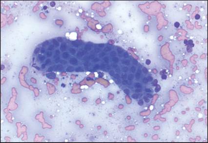
Figure 10.1 A tubule of renal epithelium (modified Wright’s, 200? magnification).
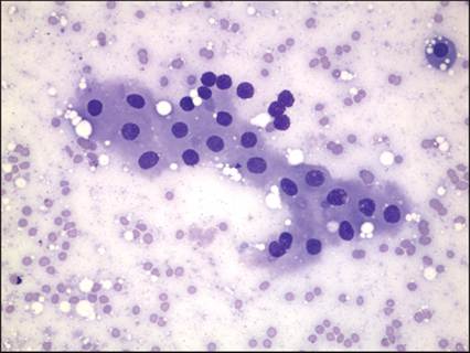
Figure 10.2 A monomorphic cluster of normal canine renal epithelium (modified Wright’s, 600? magnification).
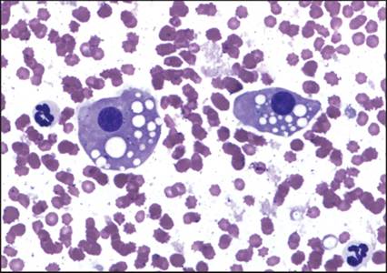
Figure 10.3 Two individualized feline renal tubular epithelial cells with many small distinct lipid droplets (modified Wright’s, 1,000? magnification).
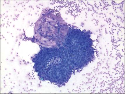
Figure 10.4 Glomerulus (modified Wright’s, 200? magnification).
More on the topic Kidneys:
- Kidneys
- There are two kidneys lying in the cranial abdominal cavity, one on each side of the midline ventral Io the lumbar hypaxial muscles (Fig. 10.1).
- Anatomy of the urinary system
- Introduction
- The Urinary System
- Kidney excretion of wastes and control of pH
- Cases
- Nephron structure
- Comparative urinary physiology and function
- If the metabolic processes of the body are to function effectively, the chemical composition and volume of the tissue Iluid must be kept constant.