Mucoid Enteropathy
Mucoid enteropathy is recognized as a major disease in domestic rabbits, but it is inappropriate to consider it a primary disease. Gastric bloat, mucous discharge, and cecal impaction, which are the cardinal features of mucoid enteropathy, are nonspecific responses of the rabbit intestine to a number of deleterious factors.
Clinical signs associated with the syndrome include bruxism, anorexia, lethargy, crouched stance, diarrhea, succus- sion splash, cecal impaction, and accumulation of large quantities of clear gelatinous mucus in the colon. Cecal impaction and mucous production occur most often in rabbits that live for 7-14 days after the onset of the syndrome. The morbidity is variable but may be high, particularly in rabbits affected during the postweaning period. There is usually a high mortality rate in affected animals regardless of the treatment. Rabbits 7-10 weeks of age are most often affected, but ages ranging from 5 weeks to adults may be involved in outbreaks of mucoid enteropathy.Many theories have been proposed to explain the etiopathogenesis of mucoid enteropathy. Dietary factors have frequently been implicated. This condition was relatively uncommon prior to the feeding of high-energy commercial rations, and rabbits fed a high-carbohy- drate/low-fiber diet have been shown to have a higher incidence of the syndrome than those fed diets high in fiber. Studies of the cecal microbial flora have revealed striking changes in rabbits with mucoid enteropathy. In normal animals, large numbers of ciliated protozoa and large, metachromatically staining bacilli are present in the cecal contents. Rabbits with mucoid enteropathy have a dramatic cecal dysbiosis. Large metachromatic bacilli and ciliated protozoa may be present in small numbers or completely absent, and there is a marked rise in coliform bacteria in the cecal contents.
Microbial instability may occur more often in young animals, where homeostatic mechanisms are poorly developed, thus increasing susceptibility to dietary changes associated with weaning.Pathology
The stomach is often distended with fluid and gas. The jejunum is frequently distended with translucent, watery fluid, and the cecum is often impacted with dried contents and gas. The colon is usually distended with characteristic clear, gelatinous mucus (Fig. 6.78). Microscopically, there is massive discharge of mucin from goblet cells in the mucosa of affected small and large intestine, with minimal or no inflammatory response. Goblet cells may be in different stages of mucin release. In the colon, crypts and the lumen of the gut are distended with mucus and mucous plugs (Fig. 6.79). Lesions are usually minimal to absent in the cecum. Mucoid enteropathy often accompanies or precedes viral and/or bacterial enteritides, or management problems that may lead to disruption of the intestinal microflora or function.
Gastric Dilation (Bloat) and Gastrointestinal Stasis
Acute gastric bloat, often accompanied by tympany of the intestine, is a life-threatening syndrome in rabbits. A number of factors are associated with bloat, including high-carbohydrate diet, dysautonomia, epizootic rabbit enteropathy (Fig. 6.33), mucoid enteropathy, and others. In 1 study, a significant majority of rabbits presenting with gastric bloat had accompanying intestinal obstruction due to bezoars, neoplasia, postsurgical adhesions, foreign bodies, tapeworm cysts, and others. Gastrointestinal stasis, including gastric and intestinal bloat, is secondary to a large number of factors, often in
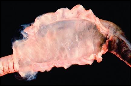
FIG. 6.78. Sacculated colon from a juvenile rabbit with mucoid enteropathy. The opened bowel is filled with clear gelatinous material.
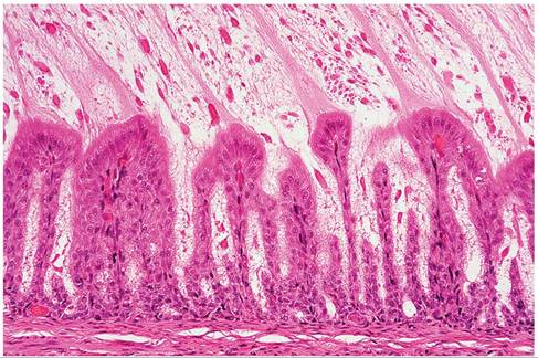
FIG. 6.79. Colon from a rabbit with acute onset of mucoid enteropathy. Note the abundant mucous within the lumen and adherent to enterocytes, depletion of goblet cells, and lack of inflammation.
combination, that cause anorexia, including dental disease, stress, infection, neoplasia, drug effects, restricted water and food intake, and so on. Once initiated, dys- motility leads to a downward spiral of abnormal colonic/ cecal transit, maldigestion, and dysbiosis. Unrelieved tympany leads to hypovolemic shock.
More on the topic Mucoid Enteropathy:
- Clostridium perfringens: Epizootic Rabbit Enteropathy
- 25 Protein losing enteropathy in a dog
- Enterococcus spp. Infection: Enterococcal Enteropathy
- Clostridium difficile and Clostridium perfringens: Clostridial Enteropathy
- Clostridium difficile, Clostridium perfringens, and Clostridium spiroforme: Clostridial Enteropathy
- Bibiliography for Noninfectious
- Intestinal Obstruction and Rupture
- Clostridium botulinum Dysautonomia: Grass Sickness
- Abbreviations list
- The Use of Corticosteroids in Otitis Media
- Flushing and Suctioning the Bulla