Salmonella enterica Infection: Salmonellosis
Salmonella, a member of the Enterobacteriaceae, continues to evoke unresolved debate over nomenclature. Among 2,500 serovars, DNA-DNA hybridization suggests that most serovars probably represent a single species, with the exception of Salmonella bongori.
The Centers for Disease Control and Prevention recognizes only two species, S. bongori and S. enterica, which is divided into 6 subspecies. Pathogenic serovars belong to S. enterica spp. enterica. The Salmonellae of significance to the mouse are S. enterica spp. enterica serovar Typhi- murium (aka S. typhimurium) and serovar Enteritidis (aka S. enteritidis). During the first half of the last century, sporadic outbreaks of salmonellosis were a relatively common occurrence in conventional colonies of mice. With improved quality control and husbandry, Salmonella infection of laboratory mice is now rare. Nevertheless, Salmonella is an extensively utilized experimental model system in mice, allowing opportunity for iatrogenic infections of mouse colonies. In addition, Salmonella infection of pet and fancy mice remains a zoonotic risk. Because Salmonellae have a broad host range, the danger of interspecies transmission, including zoonotic risk to humans, is an important consideration. Subclinical carrier animals pose a significant risk.Epizootiology and Pathogenesis
Salmonella enterica serovars Enteritidis and Typhimu- rium are the most commonly identified natural serovars in mice, and S. enterica serovar Typhimurium is a commonly used experimental serovar. Infection is initiated by ingestion of contaminated feed or bedding, although conjunctival inoculation requires fewer organisms to establish an infection. Salmonellae exist intracellularly, stimulate their own uptake by enterocytes, and continue to survive and replicate in macrophages. Host susceptibility or resistance depends on a variety of factors, including age (weanlings are more susceptible than adult mice), gut microflora, strain of mice, virulence and dose of organism, route of inoculation, intercurrent infections, and manipulations that impair the immune response.
Normal gut microflora create a natural microbial barrier to infection with Salmonella. Susceptibility to experimental infection is often enhanced by pretreatment with Streptomycin to abrogate the microbial barrier. B6, C3H/HeJ, C57BL/10ScCr, and BALB/c mice are highly susceptible, A/J and CBA/N are intermediate, and 129S6/SvEv are resistant. Resistance is mediated through several different factors, including the natural resistance- associated macrophage protein 1 (Nramp1) and TLR4, which explains the susceptibility of C3H∕HeJ and C57BL/ 10ScCr mice. The intermediate susceptibility of CBA/N mice, which have a defect in humoral immunity, can be abrogated by passive transfer of Salmonella antiserum.Following exposure by ingestion, the incubation period is usually 3-6 days. Organisms gain entry to mucosa via fimbrial attachment to M cells, followed by uptake through type III secretion systems that induce phagocytosis by enterocytes, and modification of the intracellular environment of macrophages to allow survival. Initial replication in enterocytes is followed by multiplication in gut-associated lymphoid tissue and then spread to the systemic circulation. In a small percentage of animals, there may be intermittent shedding of the organism in the feces for several months. The organism may also be harbored in the upper respiratory tract in carrier mice. Salmonella is readily killed by neutrophils, and neutrophil function is an important factor in resistance, but the bacterium has adapted to grow within macrophages, effectively evading clearance. In the liver, bacteria replicate intracellularly within macrophages, producing focal histiocytic granulomata as the hallmark lesion. Genetically susceptible mice die from massive bacterial proliferation and tissue destruction related to endotoxin.
Pathology
Clinical illness among naturally infected mice is rare, and persistently infected carrier mice are common. Clinical signs, when present, include diarrhea, anorexia, weight loss, conjunctivitis, and variable mortality.
Gross findings may include splenomegaly, with multifocal pale miliary foci present on the liver. The alimentary tract is frequently essentially normal. In other cases, there may be ileal hyperemia, and scanty fluid contents may be present in the small and large intestine. Mesenteric lymph nodes can be enlarged and reddened. There may also be scant fibrinous peritoneal exudates. Microscopic changes include multifocal necrosis and venous thrombosis with leukocytic infiltration in the liver, spleen, Peyer's patches, and mesenteric lymph nodes. The hepatic lesions are typically granulomatous in nature (Fig. 1.63). In the terminal small intestine and cecum, there may be edema in the lamina propria and submucosa, with sloughing of enterocytes and leukocytic infiltration.Diagnosis
Isolation and identification of the organism from sites such as liver, spleen, mesenteric lymph nodes, and intestine are essential and must accompany the standard macroscopic and histopathologic findings. Culture requires enrichment with Selenite F broth plus cysteine, followed by streaking on brilliant green agar. When screening a colony for possible Salmonella carriers, culturing of individual fecal samples is more sensitive than using pooled samples, and the highest rate of detection
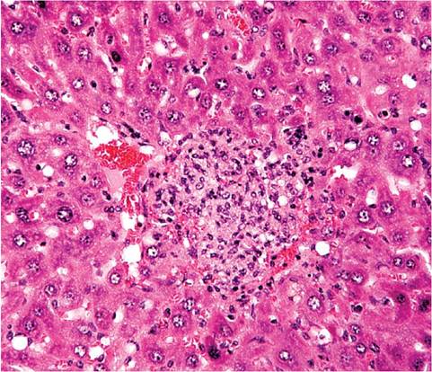
FIG. 1.63. Liver from a mouse with salmonellosis. The circumscribed lesion consists of aggregations of histiocytes.
among carriers is achieved by culturing mesenteric lymph node, since fecal shedding is intermittent. Differential diagnoses include Tyzzer's disease, coronaviral hepatitis, mousepox, Helicobacter hepatitis, and pseudo- moniasis. Spontaneous mesenteric lymphadenopathy (mesenteric disease) can also occur in aging mice.
More on the topic Salmonella enterica Infection: Salmonellosis:
- Salmonella spp. Infection: Salmonellosis
- Salmonella enterica Infection
- SALMONELLA INFECTIONS IN WILD MAMMALS
- SALMONELLA INFECTIONS IN WILD BIRDS
- Chapter 11 NOTIFIABLE DISEASES, SALMONELLOSIS AND ZOONOSES
- CHAPTER 31 SALMONELLA INFECTIONS
- Picornavirus Infection: Mouse Encephalomyelitis Virus Infection
- Arenavirus Infection: Lymphocytic Choriomeningitis Virus Infection
- Arterivirus Infection: Lactate Dehydrogenase-Elevating Virus Infection
- Coronavirus Infection: Mouse Hepatitis Virus Infection
- Streptococcus pneumoniae Infection: Pneumococcal or Diplococcal Infection
- INFLAMMATION IN HIV-1 INFECTION