the Oestrous cycle
This is the name given to the sequence of physical and hormonal events which culminate in the behavioural signs of the cow being ‘on heat’ or ‘on bulling’, or ‘in oestrus’, approximately every 3 weeks.
Puberty is the age at which an animal becomes sexually mature; that is when oestrous cycles begin. In heifers the onset of puberty can vary from as little as 6 to as much as 18 months old, with nutrition being the most important determining factor.
Physical Changes
Figure 8.2 and Plates 8.1 and 8.2 give the basic anatomy of the cow’s reproductive tract. It was also shown in detail in Plate 5.1. At birth, the ovary contains all the eggs the cow will need for her reproductive life (some 75,000 eggs are present in each ovary!) and from puberty onwards one egg is passed down into the uterus every 21 days, interrupted only by pregnancy and a short period of ovarian inactivity in early lactation. When it is ready to be shed, the egg is contained in a small fluid-filled sac on the surface of the ovary called a follicle.
At the end of oestrus the follicle bursts and releases the egg into the oviduct. This is known as ovulation (see Figure 8.3). The egg then passes down to the junction of the oviduct and uterus, and this is the point where fertilisation may take place.
Immediately after ovulation, glandular tissue begins to form in the base of the ruptured follicle and it grows until there is a mass protruding from the surface of the ovary. This structure is called the corpus luteum. It is sometimes known as the ‘yellow body’ because of its colour, or simply abbreviated as ‘the corp’. It is clearly seen in Plates 8.1 and 8.2. This structure can be felt from day 4 or 5 onwards, and it is what your vet is feeling for when he is assessing whether or not a cow is cycling. From its shape and size he will also be able to give you some idea of how many days past the previous bulling the cow is at the time of examination and this will help you to know when to watch for her next heat.
If the cow conceives, the corpus luteum remains in the ovary for the whole of pregnancy. However, if she does not conceive it decreases in size from days 16-18 onwards and this allows the devel-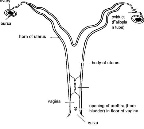
Figure 8.2. The reproductive tract of the cow.
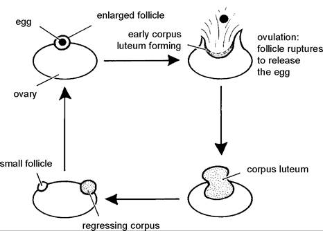
opment of a second follicle, as shown in Plate 8.2. Figure 8.3. Changes in the ovary during the oestrous cycle.
As the follicle expands and matures in preparation for ovulation, it produces increased quantities of the hormone oestrogen. It is the action of oestrogen in the body which causes the physical changes associated with oestrus including, for example, enlargement of the vulva, passage of the ‘bulling slime’ and, of course, mounting behaviour. In fact waves of follicles develop and regress throughout the oestrous cycle, with some cows having two wave cycles and others three wave cycles. A three wave cycle denotes the fact that there is increased follicular activity on the ovary on, for example, days 8, 13 and 22. A two wave cycle would have follicles at days 10 and 21 only. In each case it is only the follicles at days 20-22 which rupture to release the egg.
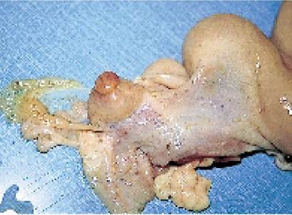
Plate 8.1. An ovary showing a corpus luteum approximately 7 days after ovulation. The convoluted tube on the left is the oviduct (fallopian tube).
Each time there is a new wave, 2-3 follicles on the ovary increase in size, a process known as recruitment. These follicles produce an increase in circulating blood oestrogen and the cow may show slightly more interest in other cows in oestrus, although it is unlikely that she will stand to be mounted. At day 21 one of the follicles ovulates and all the other follicles undergo atresia, that is they regress back to normal size and may be selected (recruitment) for another follicular wave in the future.
As one might expect, cows experiencing three wave cycles have an oestrous cycle 1 to 3 days longer than cows with two wave cycles. This helps to explain the variable cycle length in some cows. Two wave cycle cows show a better response when GnRH is used on repeat breeders (page 272).A few days after ovulation the corpus luteum begins to produce the hormone progesterone. This has almost the opposite effect to oestrogen. Progesterone suppresses the signs of heat, suppresses the release of the hormones FSH and LH from the
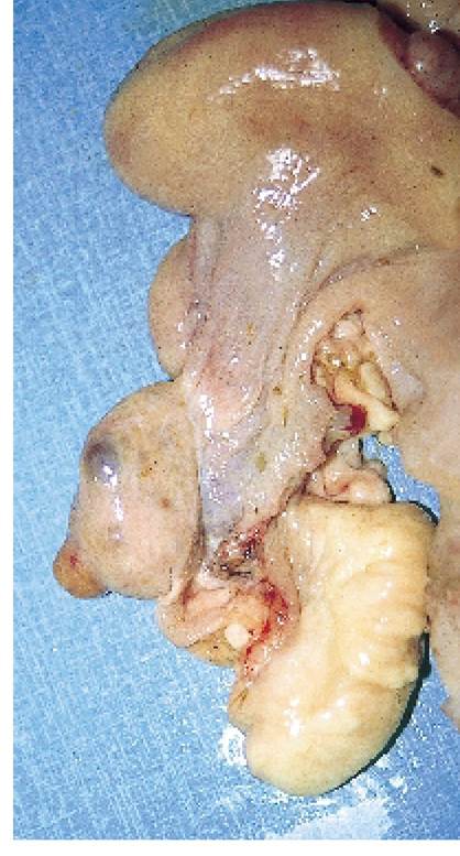
Plate 8.2. As the corpus luteum decreases in size progesterone levels fall, allowing the development and maturation of the next follicle. The plate shows an orange corpus luteum protruding from the left of the ovary and a dark grey, fluid-filled follicle in the centre.
pituitary gland (thereby inhibiting the start of the next cycle, Figure 8.4), and prepares the uterus to accept the fertilised egg, known as the ovum.
After 16 to 18 days and in the absence of pregnancy, the wall of the uterus produces the hormone prostaglandin (PG). This passes to the ovary and ‘dissolves’ the corpus luteum (Figure 8.5). Considering that it is quite a large structure, the corpus luteum regresses surprisingly rapidly: in as little as 3 or 4 days. As the corpus luteum regresses, progesterone levels fall, allowing the release of hormones from the brain to initiate the next cycle.
Hormonal Changes
The two major hormones acting on the ovary are:
follicule stimulating hormone (FSH) luteinising hormone (LH)
Both are produced and stored in the anterior pituitary gland at the base of the brain, although once produced their release is controlled by hormones from the hypothalamus, as shown in Figure 8.4. Both hormones are needed to stimulate follicular development, but LH has additional functions in that it leads to ovulation and it also promotes the growth of the corpus luteum.
Measurements of LH in the blood of a freshly calved cow show quite low activity: perhaps one ‘pulse’ is released into the bloodstream every 6-8 hours. However, as follicles get closer to maturation (under the influence of FSH) the frequency of the LH pulses increases to once every 30 minutes.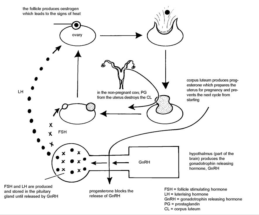
Figure 8.4. Hormonal changes during the oestrous cycle. Progesterone produced by the corpus luteum inhibits the release of GnRH from the hypothalamus. When progesterone levels fall, GnRH releases LH and FSH from the pituitary, and the next cycle starts.
This rapid pulse release is extremely important in the development of the next crop of ovarian follicles and it is interesting that it is inhibited by factors such as:
• a suckling calf (and so beef cows are slower to come on heat after calving than dairy cows)
• negative energy balance (typical of the early lactation cow at peak yield)
• progesterone from the corpus luteum
• placental steroids produced in late pregnancy
The release of LH and FSH into the bloodstream (and therefore to the ovary) is controlled by yet another hormone, namely GnRH, gonadotrophin releasing hormone. (FSH and Lh are called gonadotrophins because they influence the growth of the gonads.) GnRH is produced in the hypothalamus, and its action is inhibited by progesterone. Towards the end of a normal oestrous cycle the sequence of events, shown in Figure 8.4, is as follows:
• FSH and LH accumulate in the pituitary gland, but cannot be released because progesterone produced by the corpus luteum is blocking the action of GnRH.
• In the absence of pregnancy, the uterus produces prostaglandin around day 16.
• Prostaglandin dissolves the corpus luteum, progesterone levels fall and GnRH becomes activated.
• FSH and LH are released from the anterior pituitary gland and pass to the ovary to stimulate the development of the next crop of follicles.
Recognition of Pregnancy
In much the same way as the full-term calf tells the cow it is ready to be born (see Chapter 5), it is the embryo which sends out a signal to inform the cow that she is pregnant. This signal is a protein, known as bovine trophoblastin (bTb), and it is similar in structure to interferon. The signal inhibits production of prostaglandin by the uterus and so in the pregnant cow this leads to the following sequence of events (see Figure 8.5):
• The foetus inhibits the release of prostaglandin from the uterus.
• The corpus luteum remains in the ovary and progesterone levels stay high.
• High progesterone levels inhibit the action of GnRH, thereby preventing the release of LH and FSH, so that the next ovarian cycle does not start.
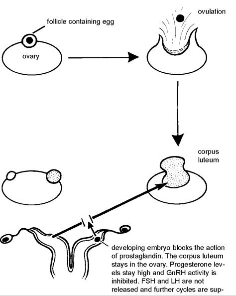
Figure 8.5. In the pregnant cow the developing embryo releases a signal (above) which inhibits uterine release of prostaglandin. In the non-pregnant cycling cow the uterus produces prostaglandin from day 16. This dissolves the corpus luteum, thereby initiating the next cycle.
If the corpus luteum is removed for some reason, for example by injecting prostaglandin or cortisone, progesterone levels fall and the cow will abort.
Action of Fertility Cycle Drugs
Many of the hormones which we have described are also available as injectable preparations and you may find it interesting to know which types of drugs your vet uses for fertility treatments.
Oestrogen
Oestrogen can be used to stimulate ovarian function in cows which have not started cycling after calving and also as a treatment for endometritis. It has two disadvantages, however. Firstly there is a danger of cystic ovaries developing after treatment and secondly the cow may only show the behavioural signs of oestrus, without going through any of the ovarian changes which lead to pregnancy.
FSH and LH
FSH and LH are commonly used to stimulate ovarian activity, or GnRH can be given to stimulate the release of FSH and LH which is naturally produced.
LH can also be used as a ‘holding injection’ on the day of service to ensure that ovulation occurs and on day 12 post service to reduce embryo death. This is described in more detail on page 272.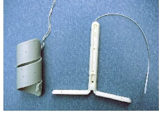
Plate 8.3. Progesterone releasing devices, a PRID (left) and CIDR (right).
Prostaglandin
Prostaglandin, either the natural hormone or a synthetic product, is an extremely commonly used drug. When given by intramuscular injection it causes the dissolution of the corpus luteum, progesterone levels fall, GnRH becomes activated, FSH and LH are released and the cow comes into oestrus 3-4 days after injection. These changes can be followed in Figure 8.4. Prostaglandin can only act if there is a corpus luteum in the ovary, however, and the corpus luteum is only sensitive to prostaglandin during days 5-15 of the cycle.
One word of caution: prostaglandin will lead to the regression of the corpus luteum whether or not the cow is pregnant, and if given to a cow at less than 150 days or more than 250 days of pregnancy, it is highly likely that she will abort. Your vet will therefore want to carry out a rectal examination of the cow prior to the administration of the drug and you should also check your records to ensure that there is no possibility of the cow having been served in the preceding 6 weeks, since pregnancies of this age or less may not be detectable by rectal examination. Prostaglandin is also used in the treatment of endometritis (page 266).
Gonadotrophin releasing hormone
GnRH is used primarily in the treatment of cystic ovaries and to improve conception rates. Its action and uses are described on pages 242 and 272.
Progesterone releasing devices
A variety of devices that maintain a continuous level of progesterone circulating in the cow’s system are on the market. This has the effect of blocking GnRH, as in Figure 8.4, and in so doing it prevents the release of LH and FSH, preventing the start of a new cycle. When the device is removed after 10-12 days, blood progesterone levels fall, GnRH becomes activated and sufficient LH and FSH will have accumulated to initiate ovarian activity, inducing a fertile oestrus two days after the device has been removed. The most common devices are shown in Plates 8.3 and 8.5.
A PRID (progesterone releasing intravaginal device) consists of a progesterone impregnated silicone rubber coating around a metal coil. The coil is inserted into the vagina, with the string protruding from the vulva (Plate 8.4) for easy removal after 12 days. The small gelatine capsule at one end (Plate 8.3) contains oestradiol. This dissolves naturally soon after insertion of the PRID and acts by removing any remaining corpus luteum. One disadvantage of the PRID is that it sometimes produces a vaginitis, with copious quantities of purulent material discharging from the vulva following its removal. However, although this looks unsightly, it does not seem to affect conception rates.
A CIDR (controlled internal drug release) consists of a nylon T- shaped spine covered by progesterone impregnated silicone (Plate 8.3) with a small plastic tail which is left protruding through the vulva. The CIDR is said to cause less vaginitis, but it has no oestradiol capsule, so prostaglandin injections are often given just prior to CIDR removal.
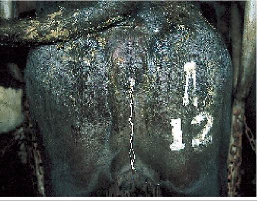
Plate 8.4. A PRID is removed after 12 days by pulling on the string, which can be seen protruding from the vulva.
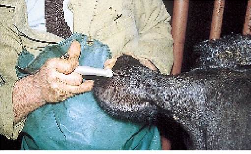
Plate 8.5. Progesterone releasing devices can also be implanted under the skin of the ear, as with this CRESTAR.
The CRESTAR is a progesterone implant which is placed under the skin of the ear, as shown in Plate 8.5. A single dose of oestradiol is given by intramuscular injection at the time of implantation and this helps to remove any remaining corpus luteum. After 9-10 days a small scalpel cut is made in the overlying skin and the implant is squeezed out. The cow comes on heat 48 hours after removal.
CRESTAR implants probably give the best heat synchronisation and as such the manufacturers state that only one insemination is required after removal, whereas for PRID and CIDR, inseminations on two consecutive days, or at observed oestrus, are recommended. There is also no risk of vaginal infections with a subcutaneous implant.
Failure of the progesterone releasing devices can be caused by:
• Persistence of a corpus luteum in the ovary. The use of oestradiol with the PRID and CRESTAR should minimise this and an injection of prostaglandin is recommended when using a CIDR.
• Insertion of the device when the cow is very close to oestrus. In a small proportion of such animals
| Event/treatment | ||
| Day | Donor | Recipients |
| 0 | Bulling | |
| 2 | Insert PRD | |
| 10 | Inject FSH/PMSG | |
| 11 | Inject FSH/PMSG | Inject prostaglandin |
| 12 | Inject FSH/PMSG | Remove PRD and inject prostaglandin |
| 14 | Observe for heat late pm | Observe and record heats |
| 15 | Inseminate Inject GnRH | Observe and record heats |
| 22 | Flush embryos | Transfer embryos |
| PRD = | a progesterone releasing device. | |
there is then a corpus luteum in the ovary when the implant is removed 10-12 days later and she fails to come on heat.
• Refractory anoestrous cows. Cows which are very thin or stressed in some other way sometimes totally fail to respond. In such extreme cases it may be better to wait until they start regaining weight before the device is administered.
If the device is left in place for significantly longer than 12 days (to allow all luteal tissue to regress naturally), the prolonged period of progesterone may depress subsequent conception rates. An alternative system, therefore, is to inject prostaglandin 1 day before the device is withdrawn, for example, inject on day 9, withdraw on day 10 (that is, a shortened period) and serve on day 12. This is obviously more expensive, but it does produce better synchronisation and more successful conception rates.
Both prostaglandin and the PRID can be used to synchronise the onset of oestrus in groups of cows or heifers, thus allowing fixed-time AI and eliminating the need for heat detection. This will be covered in more detail later in the chapter.
Embryo Transfer
Many of the drugs mentioned above are used during embryo transfer. This is a technique to increase the number of offspring from a cow of exceptional genetic merit. An embryo is an egg or ovum which has been fertilised. In the normal cow only one follicle ovulates, producing one ovum each time the cow comes on heat (possibly two for twins). However if a large dose of FSH (usually in the form of pregnant mare serum gonadotropin, PMSG) is given daily for 2-3 days before oestrus, then the number of ova shed from the ovary will be increased. This is known as superovulation. After fertilisation by AI, the resulting embryos can be flushed from the donor cow by means of catheters fed into her uterus, separated out and placed singly into recipient heifers. Table 8.1 gives a typical schedule for embryo transfer.
There are many alternatives.
The embryo is very fastidious in its requirement for uterine environment and because of this it is essential that it is transferred into a recipient which is at the same stage of the oestrous cycle as the donor. This means that the recipients must be observed to be in standing oestrus within 24 hours of the donor, and preferably less. This synchronisation of heats is achieved by using prostaglandin or progesterone releasing devices (PRDs). To ensure that the donor releases the large number of ova following superovulation, an injection of GnRH is given on the day of the insemination. Flushing of the donor to remove the Table 8.1. Atypical schedule of events for embryo transfer. The embryos from her uterus is usually time of day for some injections will be precisely specified. carried out 6-7 days after insemination. Each embryo must be carefully examined under a microscope before it is transferred into a recipient. Some eggs will not have been fertilised (that is, they are still ova and have not developed into an embryo). Some embryos may be degenerating and are not suitable for transfer. The transfer into recipients may be carried out surgically, by an incision through the flank and by depositing the embryo directly into the uterus, or non-surgically by passing a catheter through the cervix. Surgical transfer may give slightly better results but is more expensive.
What results can be expected? The great variable is the response of the donor to superovulation treatment, so the number of embryos recovered may vary between none and 25. However, an overall average result, for example, for non-surgical transfer, would be eight embryos recovered, six suitable for transfer and resulting in three established pregnancies. This may not seem a lot, but of course the donor cow is not yet pregnant. She can either be flushed again for a further crop of embryos or inseminated in the usual way. Flushing should not have any adverse effect on her subsequent fertility.
Embryos can be stored for long periods of time in liquid nitrogen. This overcomes the variable response to superovulation because once the embryos are frozen, a suitable number of recipients can be synchronised for transfer at a later date.
Cystic Ovaries
In Figure 8.3 we saw that the normal follicle ruptured to release the egg (the process of ovulation) and this was followed by the growth of the corpus luteum. Sometimes, however, instead of rupturing, the follicle continues to enlarge and this forms an ovarian cyst (see Figure 8.6 and Plate 8.6). Cystic ovaries are classically subdivided into two types, follicular and luteal cysts, depending on their development and which hormone they produce. However, this is probably an artificial subdivision, in that there is good evidence that at least a proportion of cows have cysts which intermittently produce either oestrogen or progesterone, while other cows develop cystic ovaries which resolve spontaneously. This is particularly common in early lactation. Although a cow with irregular heats should always be checked, do
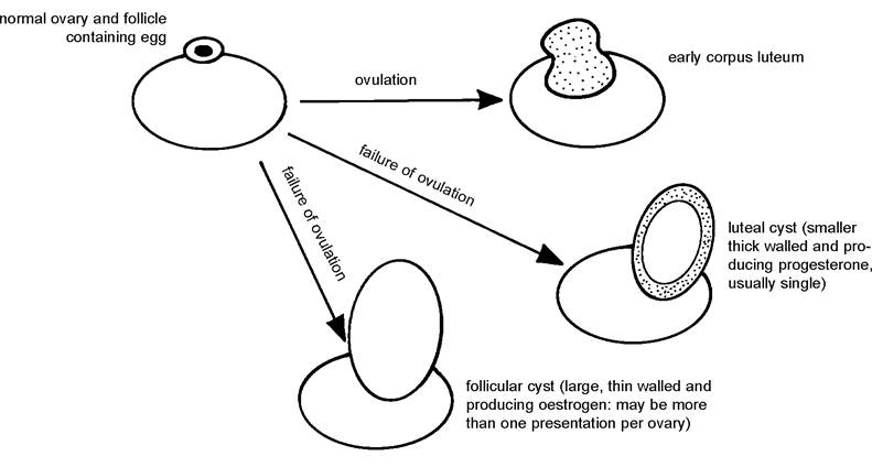
Figure 8.6. The development of cystic ovaries.
not be too surprised if no abnormalities are found.
Follicular cysts
If oestrogen is produced, the cow is said to have a follicular cyst and she shows signs of excessive oestrous behaviour. The cow is sometimes said to be nymphing, or we say that she has become a nymphomaniac. These cows will come into oestrus at irregular intervals, perhaps every 8-12 days or even more frequently, and they may stay on heat for 3-4 days instead of the normal 12-18 hours. They may also become active whenever any other cows in the herd are bulling. If left untreated, they develop a very high tail head and their pelvis may creak as they walk, due to oestrogen relaxing the supporting ligaments. Eventually masculinisation develops and the cow starts roaring and pawing the ground like a bull.
Luteal cysts
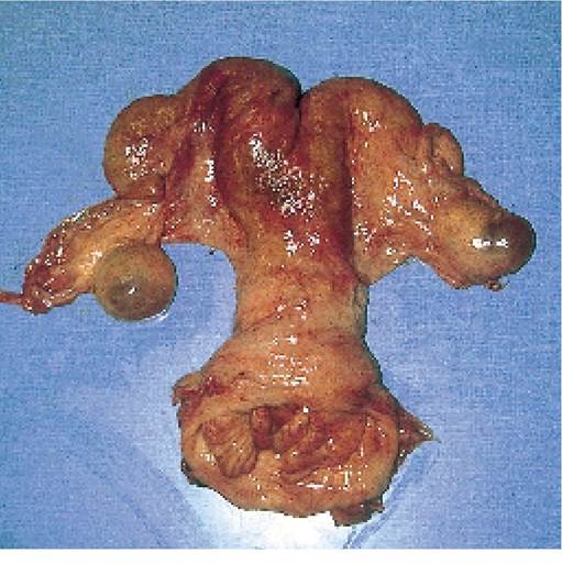
Plate 8.6. Cystic ovaries. Note the large, fluid-filled structures present in both ovaries. Some cysts can be many times larger than this.
In some cows a layer of progesterone producing luteal tissue may grow on the inside of the cyst wall and this is known as a luteal cyst (Figure 8.6). The progesterone produced by the luteal cyst blocks GnRH activity, ovarian cycles cease and the cow is never seen on heat. She is now in the true state of anoestrus, which simply means without ovarian activity.
The differentiation between follicular and luteal cysts is not always an easy matter. Only the extreme forms have been described. Intermediate stages occur and in other instances follicular cysts develop into luteal cysts which may then recover spontaneously. Differentiation may influence treatment. Follicular cysts are typically larger and have thinner walls than luteal cysts. The best diagnosis can be obtained by using milk progesterone tests (see page 244). Luteal cysts can be treated with prostaglandin; follicular cysts with Lh, GnRH or a combination of LH and progesterone. Progesterone releasing devices should be effective against both types.
Causes of cystic ovaries
Normally about 4% of cows develop cystic ovaries each year, although in some herds they can become quite a problem. The condition is partly inherited - I can remember treating a cow and two of her daughters for cysts on one farm on the same day! In Sweden, cystic ovaries once occurred in 10% of all cows, so they introduced a careful selection policy to ensure that bulls used for breeding were not derived from cows which had had cystic ovaries. This reduced their national incidence to 5%, much the same as the current level in Great Britain.
Stress is thought to be another factor involved. It causes a variety of hormonal upsets. It has been suggested that a cow under stress does not produce enough GnRH in the brain (Figure 8.4) and this leads to an inadequate release of FSH and LH. A follicle is produced and the cow comes on heat, but there is insufficient LH to cause ovulation.
Nutrition has also been suggested as a cause of cystic ovaries, particularly in high-yielding cows underfed at peak, but to my knowledge there is no direct proof of this. There have been anecdotal suggestions that cows given excess amounts of high starch concentrates immediately after calving, for example when they are offered peak intakes within the first week post-partum, may also be more susceptible to cystic ovaries. This could be related to acidosis and fatty liver, as shown in Table 6.2. Cows with fatty livers have been shown to have much higher levels of circulating prostaglandin than normal cows, and this could interfere with their oestrous cycles and subsequent fertility. Manganese deficiency may be involved, and factors such as B-carotene deficiency and the presence of certain oestrogenic toxins in the food are all possible predisposing causes.
Failure to Cycle
Most cows have started some oestrous cycle changes in their ovaries by 2-3 weeks after calving, although the first visible heat may not be seen until 4 or 5 weeks. However, a few cows remain with inactive ovaries until 60 days or more after calving and you will need to get your vet to attend to these. They are true anoestrous cows. He will carry out a rectal examination to make sure that there are no abnormalities on the ovary and then he will give a suitable treatment, most probably a progesterone releasing device. The hormones FSH and LH are responsible for the initiation of ovarian cycles and follicular development and, as described previously, the frequent pulsatile release of LH is particularly important.
Failure to cycle is most commonly seen in first calved heifers which have lost excessive bodyweight during the first few weeks of lactation - in other words, it occurs as a result of underfeeding. It is also seen in suckler cows and in this case the continued presence of the calf seems to inhibit ovarian activity. Stress, leading to increased levels of cortisone, may also be involved, since this inhibits the pulse releases of GnRH required to initiate ovarian cycles. Possible causes of stress are described in Chapter 9.
In some high-yielding cows (about 3%) ovarian cycles start but then stop again. The commonest cause of cows not seen bulling is poor heat detection, but the possibility that the cow has stopped cycling should not be overlooked and you should get your vet to check for this. The syndrome is referred to as the ‘long low progesterone’ or ‘anovulatory’ phase and its importance has been identified by means of serial milk progesterone sampling (see Figure 8.12). Sometimes such cows are said to be hovering: they may often appear to be close to bulling and show interest in other cows in oestrus, but will not stand to be mounted themselves. As such they show behavioural similarities to cows with cystic ovaries.
More on the topic the Oestrous cycle:
- While a repetitive gait cycle arises from the alternating base of support found in all bipeds, the existence of this cycle provides great opportunity for clinical and biomechanical analysis of a child with gait dysfunction.
- Phases of the Gait Cycle
- Gender, Identity, and Life-Cycle Rituals
- Light Cycle Aberrations
- Nutrients cycle at different rates according to element identity and ecosystem type
- CASH FLOW AND THE COMPANY LIFE CYCLE
- Life-Cycle Rituals
- Life Cycle Rituals
- Life-Cycle Events
- Integration and Disintegration during the Revolutionary Cycle
- LIFE CYCLE STRATEGIES AMONG PROTOZOA
- Biological and geochemical fluxes both determine the global sulfur cycle
- VIRAL LIFE CYCLE
- Life Cycle Evolution
- Definition and Cycle of the Financial Distress
- IMPAIRMENT IDENTIFICATION IS FACILITATED BY SUBDIVIDING THE GAIT CYCLE
- PROTEASE (PR) IN VIRUS LIFE CYCLE AND AIDS PATHOGENESIS