Bovine Models
16.3.1 Long-term bovine MAP challenge model
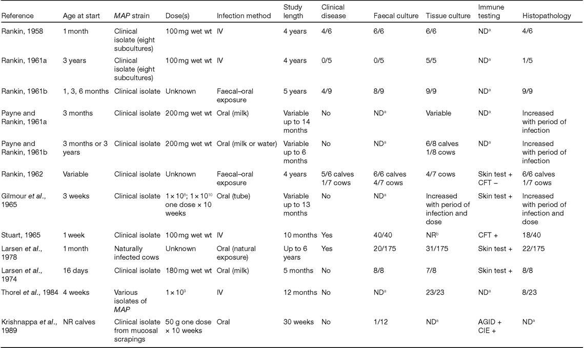
Because of the chronic nature of paratuberculosis, bovine infection models that examined inoculated animals for long periods (>6 months) are common (Table 16.3).
This long-term bovine model is typically used for studies of vaccination efficacy, preventive or therapeutic interventions, long-term pathogenesis, the immune response and assessment of diagnostic assays. A wide variety of MAP strains has been used in experimental infections (Table 16.3). Many reports identify strains only as ‘M. johnei,, ‘field isolate' or ‘clinical isolate', while others provide more specific strain designations. Strain K-10 was selected by the AMSC as the prototype strain for bovine infection models because it is known to be pathogenic and has a well-characterized genotype. However, the laboratory passage status of the K-10 MAP strain is currently unknown. Other low-passage virulent strains with a similar| Table 16.2. Main features of the murine and leporid (rabbit) models of paratuberculosis. | ||
| Model | Murine | Leporid (rabbit) |
| Breeds | C57/BL6, BALB/c, SCID C3H, Beige, Swiss Albino | White New Zealand |
| Route of administration | Oral, intraperitoneal | Oral |
| Infectious dose | 107-108 CFU/animal | 105-109 CFU/animal |
| Sample collection | Liver, spleen, intestine, lymph nodes, sera | Liver, spleen, intestine, lymph nodes, sera |
| Diarrhoea | Never | Frequent |
| Faecal shedding | Rarely | Occasionally |
| Experimental endpoints | CFU count, histopathology, immunological evaluations | CFU count, histopathology, clinical signs |
| Shortcomings | Rare development of clinical signs of paratuberculosis | Lack of reagents, incomplete clinical signs of paratuberculosis |
CFU, colony-forming units; IT, Intratonsillar; SC, Subcutaneous; ATCC, American Type Culture Collection; IFN, Interferon; LBT, Lymphocyte blastogenesis test; IL, Interleukin.
short-segment repeat genotype (e.g. 15g-5ggt genotype; Ghadiali et al. (2004)) or equivalent pulsed-field gel electrophoresis or amplified fragment-length polymorphism genotype may also be used (Hines et al., 2007b). This allows use of various local MAP strains and minimizes regulatory issues concerning the import of infectious organisms.
Types of inocula that have been administered include faeces from an infected animal, intestinal mucosal scrapings, lymph node homogenates or in vitro-cultured MAP (Table 16.3). In previous studies, MAP has been harvested from solid media (Herrold's egg yolk, Middlebrook 7H11, Middlebrook 7H10, Taylor's, Dubos) or from broth (Middlebrook 7H9). Inocula prepared from homogenized intestinal tissue, lymphoid tissue or faeces have been used, but these are difficult to standardize and aliquot for numerous studies. To address these issues, the AMSC has suggested that the chosen strain of MAP should be propagated in vitro from a master seed stock to mid-log-phase growth. Middlebrook 7H9 broth, supplemented with Oleic AlbuminDextrose Catalase (OADC), mycobactin J and 1% glycerol, was recommended as the culture medium (Hines et al., 2007b).
Storage of the inoculum at 4°C beyond 4 weeks may result in reduced viability (R. Whitlock, Philadelphia, 2008, personal communication) but storage up to 2 weeks at 4°C is generally considered acceptable. However, it is recommended that the stored inoculum should be incubated at 3 7°C for 2-3 h prior to administration. All samples within a study should be treated in the same manner.
Oral, intragastric and parenteral routes of inoculation have been successfully used (Table 16.3). However, the oral route is recommended by the AMSC as this most closely mimics natural exposure and allows MAP uptake by tonsillar tissue as well as the intestinal tract. The calf is induced to suckle from a syringe containing MAP mixed with a small volume of milk replacer or pasteurized milk. The mixture can also be gently expressed over the back of the tongue to induce swallowing.
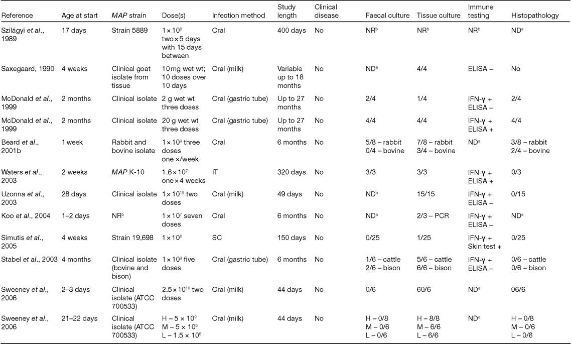
Administration by gastric tube is not recommended (Hines et al., 2007b).Published doses range from 10-200 mg wet weight and 106 to 1011 CFU (Table 16.3) but were not based on standardized procedures. Doses of 5 ? 108 CFU given on 2 consecutive days reliably resulted in infection in calves by 12-14 weeks post-inoculation (Sweeney et al., 2006). The AMSC recommends use of a standard bovine challenge dose of approximately 109 CFU/dose (100 mg wet weight) given on each of 2 successive days. Quantification of CFU should be by serial dilution and plating on solid medium known to support the organism without added antibiotics. Excessively large doses that produce clinical disease in cattle less than 18 months post-inoculation should be
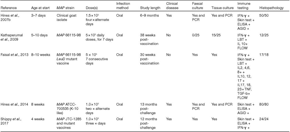
Table 16.3. Bovine models for Mycobacterium avium subsp. paratuberculosis (MAP) infection.
A.M. Talaat eta∕.
Continued
ăî σ>
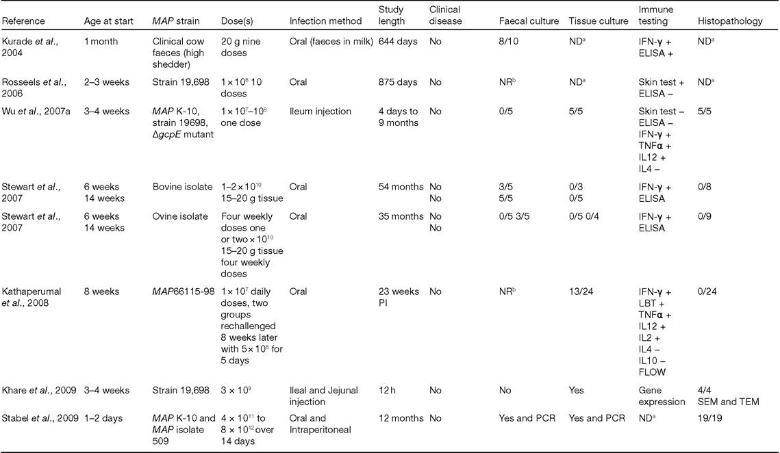
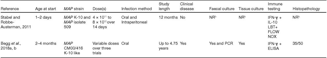
Table 16.3. Continued
A.M. Talaat eta∕.
aND = not determined.
bNR = not reported.
AGID1 agargel immunodiffusion test; CFT1CompIementfixation test; CIE1 crossed Immunoelectrophoresis; FLOW,flow cytometry; NOX1 nitric oxide; PI, post-inoculation; SEM,scanning electron microscopy; ŇĹĚ, transmission electron microscopy; ELISA, enzyme-linked immunosorbent assay; PCR, polymerase chain reaction.
avoided as they are not typical in naturally occurring paratuberculosis (Hines et al., 2007b).
Previous studies reported challenge of cattle from 1 day of age to adulthood (Table 16.3).
While studies have suggested age-associated reduction in susceptibility (Hagan, 1938; Doyle, 1953, 1956; ; Larsen et al., 19 75), a definitive relationship between infectious dose and age has not been established yet. Some flexibility in age of inoculation is required due to differences in experimental objectives and timing of other interventions, such as vaccination. Although some have suggested breed differences in susceptibility to infection (e.g. increased susceptibility of Jersey cattle to natural infection; Koets et al., 2000), this has not been formally evaluated in an experimental infection study. However, because of the difference in clinical presentation of paratuberculosis in Bos indicus breeds, the AMSC recommends that Bos taurus breeds be used exclusively for studies of bovine paratuberculosis (Hines et al., 2007b). Current experience suggests that 100 mg pelleted wet weight on 2 successive days results in demonstrable infection in calves less than 8 weeks of age (Hines et al., 2007b).Current diagnostic methods are ineffective for screening of individual calves at or before 8 weeks of age. Calves should be purchased from paratuberculosis-free farms to ensure lack of prior exposure. In the USA, herds that have achieved the equivalent of Status Level 3 or 4 in the National Voluntary Bovine Johne's Disease Status Control Program should be chosen as source herds (Hines et al., 2007b; Usda-Aphis, 2010). This equates to a closed herd with no history of paratuberculosis in the previous 5 years, tested annually with at least one whole-herd negative serological test and one whole-herd negative faecal test (second lactation and older animals).
Calves should have received adequate quantities of colostrum. Milk replacer, if used, should be high quality and of animal origin (i.e. casein not soy protein) and high-quality rations should be fed. Routine vaccinations may be administered but should not be given on the same days as paratuberculosis vaccination or MAP administration.
Anthelmintics and parasiticides should be given to all animals at normal dosages. If individual therapy is necessary (i.e. respiratory infection), an antibiotic known to have minimal effects on MAP (i.e. ceftiofur) should be used.Passive (pass-through) shedding occurs as early as 12 h after oral inoculation. Detection of passive shedding by culture provides additional confirmation of inoculum viability and the sensitivity of the faecal culture method. Positive faecal culture results more than 14 days after inoculation should be considered shedding due to infection, except in very heavily contaminated environments (R. Whitlock, Philadelphia, 2008, personal communication). The AMSC recommends that animals should have faecal cultures at least monthly during the course of the study. Gross examination and culture of tissues for MAP as well as histopathological examination of tissues to identify acid-fast organisms and lesions characteristic of paratuberculosis should be performed in all studies. At the inoculation dose recommended, colonization of tissues can be detected by culture in most animals by 4-12 weeks after inoculation, although culture of multiple tissues (minimum of three ileum including ileocaecal valve, three jejunum, one duodenum, one spiral colon and three mesenteric lymph nodes including ileocaecal nodes) is necessary. Although tissue samples should be culture-positive by 12 weeks, investigators should not always expect to find histological lesions at this early stage. A necropsy and histopathology scoring system should be used (Gonzalez etal., 2005; Hines etal., 2007b). The clinical status of the study animals should, at a minimum, be assessed and recorded monthly and at necropsy (Hines et al., 2007b).
The method of culturing faecal and tissue samples should permit quantification (or at least semi-quantification, e.g. by counting CFU on solid medium or time to positive detection in automated liquid culture systems). Decontamination by incubation of faecal and tissue samples in 0.6% hexadecyl pyridinium chloride (HPC) for 14-16 h and 3 h, respectively, is strongly recommended (Hines et al., 2007b).
There is wide variation in culture methods currently used and batch-to-batch differences occur in media. All samples should be processed on medium from the same batch. Fresh samples may be processed immediately or frozen at -70°C and thawed only once, with all samples treated in the same manner.16.3.2 Short-term bovine MAP challenge models
Experimental MAP infection models that employ direct surgical access to the ileum for administration of MAP and collection of intestinal samples have been described (Allen et al., 2005, 2009). In general, these models are most useful for short-term study of host-pathogen interactions. The time frame of such studies can vary from a few hours to a few days or even a few weeks (Wu et al., 2007a). Many parameters should be the same as for long-term challenge, including strain, inoculum preparation and quantification, storage, animal selection criteria and quality control issues. Exceptions are age of administration, dose, experimental endpoints and sample collection.
Ileal cannulation model
In this model, calves are cannulated at 8 weeks of age under general anaesthesia, using a modification of the method of Streeter et al. (1991), as previously described (Hines et al., 2007b; Allen et al., 2009). Calves are inoculated in the ileum with 1010 CFU of MAP in 20 ml phosphate-buffered saline. MAP is taken up by M cells and by dendritic and epithelial cells of the ileum and jejunum within 30 min (Momotani et al., 1988; SigurSardottir et al., 1999, 2001). The inoculation procedure is repeated the next day. Ileal mucosal biopsies can be obtained at various time intervals, to test for MAP infection and local immune responses.
Invasion/surgical model
Surgical incision and direct deposition of MAP into the ileum has been recently employed to establish a model for intestinal invasion to other organs within a few hours of infection (Wu et al., 2007a). Although infected calves survive the surgery and repeated biopsy sampling up to 10 months following infection, the focus of this model is to examine early MAP intestinal interactions. The movement of MAP from intestine to liver, spleen or mesenteric lymph node was shown to differentiate between MAP strains with different virulence phenotypes.
Intestinal loop model
An alternative surgical approach is the ligated intestinal loop model with an injected inoculum of 3 ? 109 CFU (~300mg wet weight) (Momotani et al., 1988; Khare et al., 2009). This is only suitable for studies related to bovine strain K-10, is recommended (Hines et al., 2007b).
The recommended method of inoculum preparation, quantification and storage is the same as stated above for the bovine model. The majority of studies used Middlebrook 7H9 + OADC + mycobactin J + glycerol or Tween 80 for in vitro cultivation. This medium with the addition of glycerol (1%), but without Tween or antibiotics, is considered to be the best choice (Hines et al., 2007b). The routes of administration have varied (Table 16.4), and essentially all routes tested have been successful in establishing infection, with even the aerosol route resulting in intestinal pathology (Harding, 1957). The oral route most closely parallels natural exposure and is generally considered the best route of administration.
The size of the challenge inoculum has also varied widely (Table 16.4), ranging from 2.3 7 to 200 mg and 3 ? 107 to 8 ? 1010 CFU, and all were generally successful in establishing infection. Two consecutive daily doses of 109 organisms (approximately 100 mg pelleted wet weight/dose and ~200 mg total dose) should establish infection in most kids without overwhelming experimental interventions (Hines et al., 2007b).
The age at which to inoculate has varied from the day of birth to 10 months of age (Table 16.4). It is not known whether age-related resistance occurs in goats. The age of administration will depend on the experimental goals and endpoints, but generally the goats should be less than 4 months of age. Clinical disease is expected to develop in a low percentage of animals at 9-10 months post-inoculation (Hines et al., 2007b).
The experimental endpoints will depend on the goals of the study. The minimal AMSC recommendations for goat experiments were to determine infection status through a combination of culture, PCR and histopathology, using quantitative or semi-quantitative methods (Hines et al., 2007a, b). A lesion grading system for gross and histopathological findings (Hines et al., 2007a, b) should be used, with a sufficient range in values to allow statistical analysis. Recommendations for methods of faecal and tissue MAP culture and for sample handling are the same as for the bovine model. Like cattle, positive faecal cultures within 2 weeks or more post-inoculation should be considered to be due to infection. Faecal cultures/ PCR should be performed at least monthly from all animals.
16.4.2 Short-term caprine MAP challenge models
In general, these models are used for short-term study of host-pathogen interactions. Many parameters should be the same as for long-term challenge, including strain, inoculum preparation and quantification, storage, animal selection criteria and quality control issues.
Intestinal loop model
An intestinal loop model, as previously described in goats by SigurSardottir et al. (2001), can be used for studying initial paratuberculosis bacterial-host interactions including bacterial attachment and internalization, bacterial localization, cytokine regulation and early bacterial gene regulation. MAP strain, dose, quantification, culture medium, animal selection and sample collection should be similar to the long-term challenge model (Hines et al., 2007b). Strain selection and quantification, animal selection and sample collection should also be similar to those for the long-term challenge model.
Table 16.4. Caprine models for Mycobacterium avium subsp. paratuberculosis (MAP) infection.
| Reference | Age at start | MAP strain | Dose(s) | Infection method | Study length | Clinical disease | Faecal culture | Tissue culture | Immune testing | Histopathology |
| Harding, 1957 | NRa | NRa | NRa | IH IV IV/oral oral | 9-16 months | NRa | NDb | 17/24 | NDb | 23/24 |
| Van Kruiningen et al., 1986 | 2-12 days | Human isolate Linda | 3.2?107 4.0?108 50 mg wst wt | Oral (milk) | Upto 310 days | No | 1/4 | 4/4 | NDb | 4/4 |
| SigurSardottir et al., 1999 | 7-26 days | Clinical goat isolate (three passages) | 10mg dry wt 1 ? 10 days | Oral (milk) | Up to 49 weeks | No | 0/8 | 2/8 | Skin test + ELISA + CFT + | 3/8 |
| SigurSardottir etal., 2001 | 18-21 days | Clinical goat isolate | 2.365 mg dry wt∕3ml four loops | Distal ileal ligation | 1 h | No | NDb | NDb | NDb | MAP in M cells and leucocytes |
| SigurSardottir etal., 2001 | 23-39 days | ATCC bovine strain 19,698 | Three? 107 per sleeve | Everted intestine sleeve | 1 h | No | NDb | NDb | NDb | MAP uptake by M cells and enterocytes |
| Storset et al., 2001 | 5-8 weeks | Clinical goat isolate (P173) two passages | 10mg three? 1 0 weeks | Oral (milk) | Upto 117 weeks | Yes | 4/7 | 5/7 | IFN-γ + LBT + ELISA + | 6/7 |
| Valheim et al., 2002 | 5-8 weeks | Clinical goat isolate (P173) two passages | 10mg three? 1 0 weeks | Oral (milk) | Upto 117 weeks | Yes | NDb | 5/7 | NDb | 6/7 |
| Munjal et al., 2005 | 5-8 weeks | Clinical goat isolate (tissue) | 1 ?101° seven ? 2 days | Oral | Upto 270 days | Yes | 1/10 | 2/10 (PCR) | LBT + ELISA + AGID + | 5/10 |
| Stewart et al., 2006 | 5 months | Clinical bovine isolate (tissue/ culture) | 1 ?101° 20 g wet wt | Oral | 54 months | Yes | 10/10 | 8 | IFN-γ + ELISA + | NRa |
| Stewart et al., 2006 | 10 months | Clinical sheep isolate (tissue/ | One? 4 weeks | Oral | bgcolor=white>35 monthsYes | 9/10 | 1 | IFN-γ + ELISA + | NRa |
culture)
A.M. Talaat et al.
aNR = not reported.
bND = not determined.
ELISA, enzyme-linked immunosorbent assay; PCR1 polymerase chain reaction; TNF1 tumour necrosis factor; TGF1 tumour growth factor; CFU1 colony-forming units; AGID1 agar gel immunodiffusion test; OFT, complement fixation test; LBT1 lymphocyte blastogenesis test; ATCC, American Type Culture Collection; IFN, Interferon; LBT, Lymphocyte blastogenesis test; FLOW, Flow cytometry; IL, Interleukin.
Everted intestine sleeve model
An everted intestine sleeve model, as described by SigurSardottir et al. (2005), is also useful for studying initial host-bacterial interactions, such as bacterial attachment and internalization, bacterial localization and early bacterial gene regulation, but may have limitations when evaluating host gene and early cytokine regulation in response to infection. Approximately 1.0-cm segments of small intestine are excised, everted, washed, maintained in tissue culture and bathed in a suspension of MAP for short periods of time. Strain selection and quantification, animal selection and sample collection should be similar to those for the longterm challenge model.
16.5
More on the topic Bovine Models:
- Bovine Tuberculosis
- BOVINE VIRAL DIARRHOEA
- Bovine Tuberculosis in Zambian Wildlife
- Bovine Tuberculosis Control Programs in South Africa
- Bovine TB Eradication Scheme
- Rodent Models
- Comparative Genotyping of Human and Bovine-Derived MAP Isolates
- Vaccine Models
- 8.3 Bovine Tuberculosis in African Cattle Populations
- 3. Models and definitions
- Cervid Models
- Competition in other 2-consumer-l-resource models
- Bovine TB in Ethiopia
- Lottery and neutral models rely on equality and chance
- A brief guide to externalities in growth models